


The equine heart is a remarkable organ that supports a horse’s athletic performance, endurance, and overall health. However, like any system, it can be affected by disease or structural abnormalities that compromise its function.
Detecting heart problems in horses can be challenging because many conditions develop silently, showing subtle or no outward signs until performance declines or clinical symptoms appear. This is where echocardiography plays a crucial role in equine veterinary medicine.
An echocardiogram, often called an “echo,” allows veterinarians to visualize the heart in real time and assess both its structure and function. Unlike other diagnostic tools, such as auscultation (listening with a stethoscope) or electrocardiography (ECG), echocardiography provides direct images of the heart’s chambers, valves, and blood flow patterns.
Read on to learn how echocardiograms are performed in horses, what they reveal, and how veterinarians use the results. By understanding this diagnostic test, horse owners and caretakers can better appreciate its importance in protecting equine health and performance.
Echocardiography for Horses
Echocardiography is the process of using an ultrasound to examine the heart’s function and structure. Careful evaluation of the heart allows veterinarians to diagnose heart disease and determine its severity. [1]
Also known as a cardiac ultrasound, echocardiography is non-invasive and safe for horses, with no associated complications.
Your veterinarian may recommend echocardiography for your horse for a number of reasons, including: [1][2]
- A heart murmur audible on auscultation (listening with a stethoscope)
- Arrhythmias identified on an electrocardiogram or on auscultation
- Muffled heart sounds on auscultation
- Fever of an unknown origin
- Horses with poor athletic performance or exercise intolerance
- Horses with weakness or collapse
In some cases, an echocardiogram may be a component of a pre-purchase examination. [3] The information gained from an echocardiogram helps potential buyers decide whether they want to purchase a horse with a potential cardiac defect. [3]
What an Echocardiogram Evaluates
A comprehensive echocardiogram evaluates multiple aspects of the heart, including: [2]
- Abnormal movement of the heart
- Size of the heart chambers and vessels
- Function of the heart valves
- Disturbances to normal blood flow
- Blood flow volume and pressure estimates
- Physical abnormalities with the heart
What's your top priority with your horse's health?
Enter your email to receive your store credit
Equipment
The main piece of equipment required for an echocardiogram is an ultrasound machine. This machine emits ultrasound waves through a transducer (also called a probe). [4]
The surface of the transducer contains piezoelectrics, ceramic crystals that produce soundwaves when electricity runs through them. [4] The piezoelectrics can also do the reverse, producing electricity in response to a soundwave. [4]
Electricity running through the piezoelectrics emits a soundwave, which transmits through the body’s tissues. As the soundwave encounters tissue, it bounces off and reflects back towards the transducer.
The piezoelectrics convert this wave into an electrical signal, which is interpreted by the ultrasound machine as an image. [4] This image is displayed on the ultrasound machine for interpretation by the veterinarian.
Each tissue type has its own level of echogenicity, or density on an ultrasound scan. This leads to variation in the way each tissue appears on the echocardiogram. [1]
Substances like air and bone reflect soundwaves easily, producing a bright white area on the image. [1] Fluids, including blood, absorb soundwaves and appear black. [1] Most tissues, like muscle, have an intermediate sound reflection property, which makes them appear gray.
Ultrasound machines can also measure blood flow velocity in Doppler mode. This setting measures the difference between the frequency of the soundwave emitted and the frequency of the soundwave received. [1] This allows it to calculate the speed of particles, such as red blood cells, flowing within the heart. [1]
On the ultrasound screen, this speed is typically visualized as a color array superimposed over the standard ultrasound image. [1]
Other equipment necessary for an equine echocardiogram includes: [1]
- Stocks or other restraint
- Ultrasound gel
- Sedation in some cases
Procedure
Echocardiograms of horses require precise placement of the ultrasound probe to evaluate the cardiac structures. Veterinarians typically perform echocardiography in-clinic where they have stocks or other restraint equipment available to keep the horse still during examination. [1]
In general, veterinarians avoid sedating horses for echocardiograms as sedation can affect some heart function measurements. [1] However, fractious or nervous horses can be sedated if necessary.
The procedure begins by clipping the horse’s axilla (armpit regions) and cleaning the skin surface. Once the horse is clean, the veterinarian applies ultrasound gel to the area to ensure effective conduction between the ultrasound probe and the skin. [1]
After preparing the skin, the veterinarian can begin their examination. They place the probe between the ribs to avoid reflection of the ultrasound wave by bone. [2]
They examine several locations on both the right and left axilla of the horse to ensure a complete assessment. They can use Doppler or other specialized ultrasound modes to evaluate blood flow and pressure. [2]
After completing the examination, the ultrasound gel is wiped away and the horse is ready to return home or to their stall.
Interpreting Results
The heart is a complex structure requiring careful evaluation on echocardiogram. The veterinarian interprets these results and compares your horse’s measurements with reference intervals (values considered normal) to make a diagnosis.
The heart consists of four chambers lined by muscular walls. The right side of the heart primarily handles oxygen-poor blood and delivers it to the lungs for oxygenation. The left side receives oxygen-rich blood from the lungs and distributes it to the rest of the body. [1]
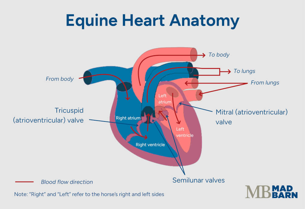
Each side of the heart has a chamber at the top called an atrium. These chambers receive low-pressure blood from the horse’s veins. Ventricles sit beneath the atria on both sides. These large chambers deliver blood into the arteries for distribution to the lungs or body systems. [1]
During a heartbeat, the atria contract to push blood through the atrioventricular valves into the ventricles. Once blood is in the ventricle, the valve closes and the ventricular walls contract rapidly.
This pressurizes the blood within the ventricle, pumping it into the arteries under high pressure. Semilunar valves help prevent blood from flowing back into the ventricles once it leaves. [1]
Heart disease can affect several aspects of this highly coordinated system. If there is too much blood in one chamber, then the wall of that chamber can stretch and become less effective at blood delivery. If there is an obstruction to blood flow, the chamber walls may thicken as they try to force blood past the obstruction. These are the changes that veterinarians try to identify on an echocardiogram. [2]
Some common conditions of the equine heart identifiable on echocardiogram include:
- Mitral regurgitation
- Aortic valvular regurgitation
- Tricuspid regurgitation
- Ventricular septal defect
- Patent ductus arteriosus
Mitral Regurgitation
The mitral valve is the atrioventricular valve between the left atria and ventricle. This valve has an important role in keeping blood within the ventricle from re-entering the atria during the powerful contraction of the ventricular walls. Regurgitation occurs when the valve becomes leaky due to structural damage.
Around 3% of horses have mitral regurgitation. [1] It typically results from wear and tear on the valve that occurs during high intensity exercise. It can also occur if there are structural problems elsewhere in the heart, such as congenital malformations. [1]
Horses with mitral regurgitation have a high risk of developing atrial fibrillation. [3] As the mitral valve leaks, it allows blood to flow back into the atria from the ventricles. This overloads the atria’s capacity, causing the walls to stretch.
Over time, this can predispose the atrial walls to abnormal electrical conduction. [3] This may impact the horse’s performance and eventually lead to congestive heart failure.
Signs of mitral regurgitation on echocardiogram include: [1]
- Prolapse of the valve (abnormal movement)
- Nodules or thickening of the valve leaflets
- Enlarged left atrium or left ventricle
- Abnormal blood flow through the mitral valve
- Enlarged pulmonary arteries (vessels that carry blood to the lungs)
Aortic Valvular Regurgitation
The aortic valve prevents backflow of the high-pressure arterial blood into the heart. Regurgitation in this valve is very common, with studies showing up to 5-9% of horses have this form of valvular insufficiency. [1]
It is most common in older horses, as it typically results from wear and tear on the valve over time. [1]
Horses with mild aortic regurgitation can have performance careers and a normal life expectancy. [3] Severe disease can rapidly result in atrial fibrillation or congestive heart failure, which increases the risk of sudden death. [3]
Echocardiogram findings related to aortic regurgitation include: [1]
- Prolapse of the valve
- Enlarged left ventricle with a thin ventricular wall
- Abnormal blood flow through the valve
- Nodules or thickening of the valve leaflets
- Poor fractional shortening (ability of the ventricle to completely empty during a heartbeat)
Tricuspid Regurgitation
The tricuspid valve is the atrioventricular valve sitting between the right atria and ventricle. The cause of tricuspid regurgitation is unknown but may be due to excessive wear from high intensity exercise. [1]
Many horses can have successful performance careers despite tricuspid regurgitation. [1]
Findings on echocardiogram related to tricuspid regurgitation include: [1]
- Prolapse of the valve
- Nodules or thickening of the valve leaflets
- Enlarged right atrium
- Abnormal blood flow through the tricuspid valve
Ventricular Septal Defect
Ventricular septal defect (VSD) is the most common congenital heart abnormality in horses. [1] This abnormality results in a hole in the interventricular septum, the muscular wall that separates the two ventricles.
In cases of VSD, blood flows from the high-pressure left ventricle into the right ventricle during a heartbeat, causing excess blood volume in the right ventricle. [1]
Horses with small VSDs can have a normal life expectancy and performance capability. Large VSDs can cause congestive heart failure and carry a poor prognosis. [3]
Echocardiogram findings related to VSD include: [1]
- A hole or defect in the interventricular septum
- Blood flow across the interventricular septum during a heartbeat
- Enlarged left and right ventricles
Patent Ductus Arteriosus (PDA)
The ductus arteriosus is a normal connection that exists between the aorta and the pulmonary arteries during fetal development. [1] After birth, this connection closes to allow normal cardiac function. In some cases, this connection remains open and allows aortic blood to leak into the pulmonary arteries. [1]
This increases the amount of blood returning from the lungs dramatically, causing high blood volume in the left atria and ventricles and stretching of the muscular walls. [1]
Very small PDAs may cause no issues for the horse. [1] However, larger openings with abundant blood flow eventually lead to congestive heart failure and potentially sudden death. [1] Ongoing examinations are necessary to monitor the progression of disease for horses with PDA.
Findings associated with PDA on echocardiogram include: [1]
- Blood flow between the aorta and pulmonary arteries
- Retrograde (opposite to the normal direction) blood flow in the pulmonary arteries
- Enlarged left ventricles
- Poor fractional shortening

Preparing Your Horse for Echocardiogram
Echocardiograms are often a specialty diagnostic procedure performed by referral equine veterinary clinics. It’s often necessary to bring your horse to these clinics to ensure an ideal environment for evaluation.
Otherwise, there is no special preparation necessary for an echocardiogram.
If your horse is nervous about clipping, you may want to practice clipping prior to the appointment or have your horse sedated and clipped several hours in advance.
Allowing the sedation to wear off completely before the examination helps ensure accurate measurements. [2]
Complications
Echocardiogram is a non-invasive procedure with no reported complications. Horses may experience “clipper burn” or skin irritation due to clipping for the procedure.
Frequently Asked Questions
Here are some frequently asked questions about echocardiography for horses:
An echocardiogram, or "echo," is an ultrasound examination of the heart for horses. It uses sound waves to create real-time images of the heart's chambers, valves, and blood flow.
Your veterinarian may recommend an echocardiogram if your horse has a heart murmur, irregular heartbeat, poor performance, unexplained collapse or weakness, or as a follow-up after other cardiac tests (such as an ECG).
The procedure is non-invasive and usually done while the horse is standing. A small area of hair is clipped behind the elbow, ultrasound gel is applied, and the probe is placed against the chest wall to capture images of the heart. Sedation is rarely required.
No, echocardiography is not painful for the horse. Most horses stand quietly throughout the procedure.
A standard echocardiogram usually takes 30–60 minutes, depending on the horse and the complexity of the findings.
Typically, no fasting or medication is needed before an equine echocardiogram. Your veterinarian may recommend clipping a small patch of hair to ensure a high-quality image is captured.
Summary
An echocardiogram is a non-invasive ultrasound test that helps veterinarians assess the structure and function of a horse’s heart.
- It is used to investigate heart murmurs, arrhythmias, poor performance, or unexplained collapse.
- The procedure is performed while the horse is standing, usually without the need for sedation.
- An echocardiogram can detect valve disease, chamber enlargement, congenital defects, and blood flow abnormalities.
- The examination typically takes about 20 - 30 minutes and requires minimal preparation.
- The results help guide management, monitoring, and long-term care decisions for the horse.
References
- Marr. C. M. and Bowen. I. M., Eds. Cardiology of the Horse. 2nd ed. Saunders, Edinburgh; New York. 2010.
- Schwarzwald. C. C. Equine Echocardiography. Veterinary Clinics of North America: Equine Practice. 2019.
- Reef. V. B. Assessment of the Cardiovascular System in Horses During Prepurchase and Insurance Examinations. Veterinary Clinics of North America: Equine Practice. 2019.
- Ultrasound. National Institute of Biomedical Imaging and Bioengineering.


