


Eye prolapse, also known as proptosis, is one of the most severe eye injuries that can occur in horses. In this condition, the entire eyeball (globe) is displaced from its normal position within the eye socket.
This injury is usually the result of trauma, such as a kick from another horse, a collision, or an accident involving fences or stall structures. In some cases, underlying disease such as a retrobulbar mass or abscess can contribute to the eye being forced outward.
A prolapsed eye is extremely painful and distressing to witness. The affected eye may appear swollen, protruding, or misshapen, and the horse may show signs of discomfort such as head shaking, rubbing the face, or avoiding light. Because the optic nerve and surrounding tissues are easily damaged, immediate veterinary attention is critical to relieve pain and, when possible, preserve vision.
Understanding the signs, causes, and treatment options for eye prolapse helps horse owners act quickly in an emergency, improving the chances of a positive outcome. Horses rely heavily on their eyesight to navigate and interpret their surroundings, so even minor eye injuries can have serious consequences if left untreated.
Eye Prolapse (Proptosis) in Horses
A horse’s eyes are large, delicate structures that play a vital role in helping them navigate their environment. Their prominent position on the head and limited natural protection make them especially vulnerable to injury.
Eye prolapse, also called proptosis, occurs when one of the horse’s eyeballs becomes displaced from its normal position. This can stretch or damage the optic nerve, putting the horse’s vision at serious risk.
Even a partial prolapse can threaten vision, while a complete displacement may result in permanent blindness even when treated promptly. [1]
Equine Vision
Understanding how horses see helps owners notice subtle changes in behavior, recognize potential eye injuries early, and respond quickly to protect their horse’s vision.
The horse’s sense of vision is highly specialized and reflects their status as prey animals. Their eyes are positioned on the sides of the head, giving them a wide field of view. Most of their vision is monocular, meaning each eye sees separately. Only a small area in front of them is binocular, where both eyes focus together to judge distance. [2][3]
This side placement makes horses highly sensitive to movement, helping them spot potential threats quickly. However, it also creates blind spots directly in front and behind, which increases the risk of accidental bumps or injuries. [4]
Because of these adaptations, horses can be easily startled, and their exposed eyes are more at risk during everyday activities.
Anatomy of the Equine Eye
The horse’s eye is one of the largest among land mammals and sits on the side of the head. This placement provides a wide field of vision but leaves the eye exposed to trauma. [5]
Key structures of the horse eye include: [2][6][7][8]
- Cornea: The clear front layer that lets light into the eye
- Sclera: The firm, white outer shell that protects the eye
- Iris: The colored part of the eye which controls the width of the pupil in response to light
- Pupil: A hole in the center of the iris that allows light to pass into the eye
- Lens: Focuses the light onto the back of the eye
- Retina: Contains cells that turn light into electrical signals for the brain to interpret
- Optic nerve: Carries light signals from the retina to the brain
 Illustration: Dr. Ana Mesa, PhD
 Protective Features of the Eye
The following structures shield and protect the horse’s eye: [9]
- Eyelids
- Eyelashes
- Conjunctiva – a thin, clear membrane that lines the eye
- The lacrimal system (tear production)
While these features help reduce minor injuries, they provide limited defense against severe trauma. [9]
What's your top priority with your horse's health?
Enter your email to receive your store credit
Risks Associated with Eye Prolapse
Horses see when light enters the cornea, passes through the lens, and reaches the retina, where it is converted into electrical signals that travel along the optic nerve to the brain. Any injury, especially prolapse, can disrupt this process.

“Eye prolapse is a critical emergency that requires immediate veterinary attention. The sooner the eye is evaluated and stabilized, the better the chance of preserving the eye and comfort for the horse”
— Dr. Jennifer Skaggs, DVMEquine Veterinarian
Prolapse can severely damage the delicate structures of the horse’s eye. Possible consequences include: [1][7]
- Optic nerve damage: The optic nerve is the only pathway between the eye and the brain. In a prolapse, the nerve may be stretched, compressed, or torn. Even minor stretching can cause blind spots or reduced vision, while severe damage usually results in permanent blindness in the affected eye. Once destroyed, optic nerve tissue cannot regenerate.
- Corneal exposure: A displaced or bulging eye leaves the cornea unprotected, raising the risk of ulcers, scratches, and infection.
- Swelling and inflammation: Trauma triggers swelling that can further compress the optic nerve and surrounding tissues, compounding vision loss.
Even a partial prolapse can compromise sight, while complete prolapse is usually a surgical emergency. Quick recognition and immediate veterinary care are essential for reducing complications.
Signs of Eye Prolapse
Eye prolapse occurs when the eyeball is pushed out of its normal position in the socket. This is a serious injury that can quickly threaten a horse’s vision. Even with quick veterinary care, prognosis for return of vision is guarded.
This condition can vary in severity. In a partial prolapse, the globe is pushed partially forwards while still remaining partially within the eye socket. Some structures may remain protected, and vision may be partially preserved.
In a complete prolapse, the entire globe (eyeball) is displaced, often stretching or damaging the optic nerve, which can result in permanent vision loss.
Even minor cases require urgent attention. Early recognition of warning signs is key to preserving comfort and vision.
Common signs of eye prolapse in horses include: [10][11][12]
- A bulging or protruding eye
- Swelling or redness around the eye
- Bleeding from the eye or eyelids
- Signs of discomfort or pain, such as head shaking or rubbing
- Difficulty seeing or navigating
Prompt veterinary care is essential. Early treatment increases the chances of saving the horse’s eye and prevents further complications like infection, corneal damage, or glaucoma.
Glaucoma & Eye Bulging
Glaucoma occurs when pressure inside the eye rises, damaging the retina and optic nerve. Early signs can be subtle, such as mild redness, slightly dilated pupils, or mild globe enlargement.
As pressure increases, the eye may bulge, feel firm, or show corneal changes. This bulging can sometimes be mistaken for a partial prolapse because the eye appears pushed forward. [13]
The key difference is that in cases of glaucoma, the eyeball stays within the socket, and the optic nerve is usually intact. On the other hand, a true prolapse involves displacement of the eye and potential stretching of the optic nerve. [5]
Veterinary assessment, including measuring intraocular pressure, is essential to distinguish glaucoma from prolapse and ensure appropriate treatment.
Causes of Eye Prolapse
In horses, eye prolapse is almost always linked to trauma. Because horses’ eyes are large and positioned on the sides of the head, they are more exposed than in many other animals.
Common causes of proptosis in horses include: [14][15]
- Kicks from other horses during turnout or conflict
- Head collisions with stalls, gates, fences, or trailers
- Falls or accidents while riding or handling
- Predator or environmental injuries, such as blunt trauma in pasture
In rare cases, underlying disease, such as an retrobulbar eye mass, can contribute to prolapse.
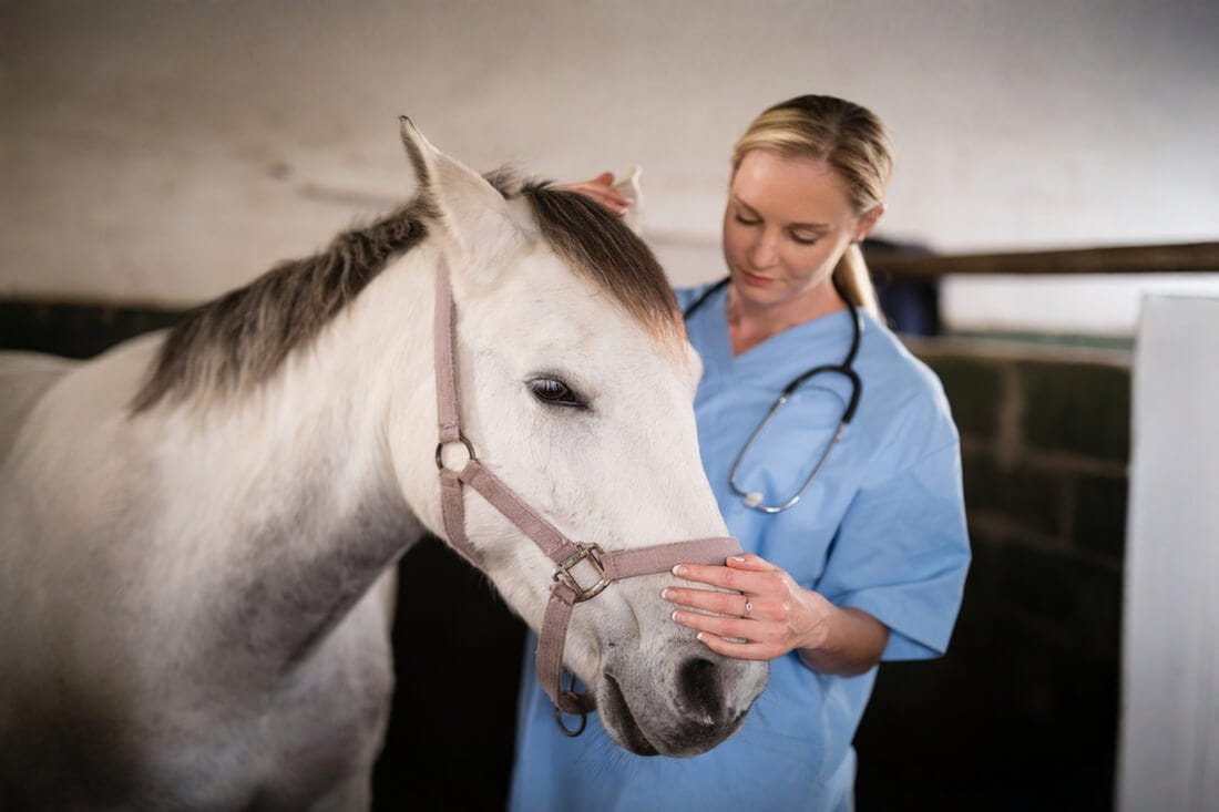
Diagnosis
When a horse shows signs of a prolapsed or severely injured eye, prompt veterinary attention is critical.
To assess a horse with suspected prolapse, the veterinarian will typically begin by observing the horse’s: [16]
- Head posture and carriage
- Facial symmetry
- Signs of discomfort or pain
- Movement through its environment, which may indicate vision impairment
Diagnosis may also involve additional tests, including: [11][14][16][17][18][19]
- Fluorescein staining: A special dye applied to the surface of the eye to reveal corneal injuries such as scratches, ulcers, or abrasions. This helps determine if the cornea is intact or at risk of further damage.
- Ophthalmic examination: Using tools like an ophthalmoscope or slit lamp, the veterinarian examines the eye’s internal structures, including the lens, anterior chamber, vitreous chamber, and retina.
- Neurological assessment: Performed if head trauma is suspected. Reflexes such as the menace response, palpebral reflex, pupillary light reaction, and vestibulo-ocular reflex are checked to detect nerve or brain damage that may impact vision.
Treatment & Management
Eye prolapse and other signs of vision changes in horses are always an emergency. Rapid treatment can make a difference in whether the eye and vision can be preserved.
The recommended treatment protocol depends on the severity of the prolapse and extent of damage to critical structures like the optic nerve. [1][14]
Treatment
The outcome of an eye prolapse depends on several factors, including: [1][12]
- Severity of the injury
- Health of the eye
- Pupil responsiveness
- How long the eye was displaced
- Presence of nearby injuries
Once the horse is stable enough for anesthesia, surgical repositioning of the eye is required. A displaced eye stretches the optic nerve and damages it, so timely correction provides the best chance of preserving vision. [1][12]
If only part of the globe (eyeball) is displaced and the optic nerve is intact, the eye is typically repositioned and temporary sutures are placed to keep the eyelids closed. Closing the eyelids temporarily protects the cornea and helps keeps the repositioned globe within the orbit until swelling recedes.
If the optic nerve or major structures are irreparably damaged, enucleation (surgical removal of the eye) is usually required to prevent ongoing pain or infection. [12]
Recovery
The management approach during recovery depends on the severity of the prolapse and any associated injuries.
The treating veterinarian will provide a tailored protocol for the individual horse, which may include a combination of: [1][12][14]
- Anti-inflammatories for pain and swelling
- Antibiotics to prevent or treat infection
- Ointments or eye drops to protect the cornea and aid healing
- Stall rest to avoid further trauma
- Controlled, supervised turnout
- Environmental changes
In cases of partial prolapse, improvement often begins within days. Swelling and discomfort usually decrease over 1–2 weeks, with full recovery taking several weeks. For a complete prolapse, the sutures and surrounding tissue may take 3–4 weeks to heal, and behavioral adjustment may take several months. [12][14]
During the recovery period, owners should watch for signs of infection or other complications including: [12][14]
- Discharge
- Redness
- Swelling
- Head shaking or rubbing
- Reluctance to move

Prognosis
The prognosis for a horse with eye prolapse depends on the extent of the injury, how quickly treatment is started, and whether secondary complications develop. [1][12]
When cases of partial prolapse are treated promptly, the prognosis can be good. Vision may be preserved, and horses can return to normal activity, including riding and turnout, once fully healed. [1][12]
Complete prolapses carry a more guarded prognosis for preserving vision. If the optic nerve or major eye structures are irreparably damaged, the eye may need to be removed. While vision in that eye is lost permanently, horses usually adjust well to life with one eye and can continue to participate in normal activities. [1][12]
Fortunately, horses can compensate for partial or complete vision loss in one eye. Initially, horses with partial blindness may be more cautious in new environments or during handling.
With supportive care and gradual exposure, confidence generally returns.
Prevention Strategies
While not every eye injury can be avoided, there are practical steps owners can take to reduce the risk of eye prolapse. Horses are prone to eye injuries because of their large, exposed eyes and natural flighty behavior, so prevention is about managing both environment and behavior.
Barn & Facility Safety
- Ensure stalls, gates, and fencing are free of sharp edges, protruding nails, or splintered wood that could injure a horse’s eye
- Use padded stall corners or panels where possible, especially for horses prone to rushing or kicking
- Maintain clear, well-lit aisles and turnout areas to reduce the risk of accidental collisions
Herd Management & Turnout
- Monitor herd interactions, particularly among new or young horses, to prevent kicks or bites
- Avoid overcrowding, as competition for space can lead to sudden movements and accidental trauma
- Consider turnout with compatible partners and separate horses with aggressive tendencies
Riding & Handling Practices
- Always be aware of surroundings, including low branches, trailer corners, or equipment that could strike a horse’s eye
- Use safe, properly fitting equipment to avoid accidental pokes or pressure near the head
- Train horses gradually in new environments to minimize spooking or sudden movements
Routine Eye Monitoring
- Inspect eyes daily for swelling, cloudiness, discharge
- Watch for behavioral signs of discomfort
- Look for subtle changes such as squinting, head tilting, or reluctance to move confidently
Early detection allows intervention before a minor abnormality becomes a prolapse. Owners should include regular inspection of their horse’s eyes as part of routine basic health checks.
Here are some tips for monitoring your horse’s eye health at home: [20]
- General appearance: Check for squinting, puffiness around the eye, and any changes in color or cloudiness within the eye. Note any alterations in eyeball shape, such as enlargement or shrinkage.
- Ocular secretions: Observe for discharge from the eyes and note the color and texture if present.
- Vision assessment: Conduct a menace response test by moving your hand toward the horse’s eye without touching the lashes or creating airflow to check for a blink or squint reaction.
| Urgency | Observation |
|---|---|
| Safe Healthy Horse |
|
| Caution Seek Veterinary Care ASAP |
|
| Urgent Seek Emergency Veterinary Care |
|
Preventing eye injuries requires a combination of environmental management, careful handling, routine monitoring, and owner awareness. While accidents can’t always be avoided, following these steps reduces the risk of serious eye injury and can help protect a horse’s vision in the long term.
Frequently Asked Questions
Here are some frequently asked questions about eye prolapse in horses:
A horse's eye can bulge due to trauma, such as kicks from other horses, collisions with stalls or fences, falls, or environmental accidents. Severe eye conditions like glaucoma can also increase pressure inside the eye, causing it to appear swollen or protruding.
Treatment for eye prolapse in horses depends on severity. Partial prolapse may be treated by repositioning the eye, closing the eyelids with temporary sutures, and using medications to control pain, swelling, and infection. Complete prolapse with optic nerve damage usually requires enucleation, or surgical removal of the eye, to prevent ongoing pain or complications. [12]
Signs of eye prolapse in horses include a bulging or protruding eye, swelling or redness, bleeding from the eye or eyelids, head shaking, rubbing, and difficulty seeing or navigating. Prompt veterinary care is essential to preserve vision and prevent further complications. [10]
Eye prolapse in horses is a veterinary emergency. Partial prolapse may allow some vision to be saved with quick treatment, but complete prolapse typically results in permanent blindness. Early recognition and rapid intervention significantly improve the prognosis.
Summary
Horses’ eyes are large, delicate, and positioned on the sides of the head, making them vulnerable to injuries such as partial or complete prolapse.
- Eye prolapse may be partial, preserving some vision, or complete, often resulting in permanent blindness.
- Common causes include kicks, collisions, falls, and environmental accidents, with diagnosis requiring prompt veterinary attention.
- Treatment ranges from repositioning the eye with supportive care to enucleation if the eye cannot be saved, followed by careful recovery and rehabilitation.
- Most horses can adapt to vision loss on one side, and many can return to riding and other normal activities.
- Prevention focuses on maintaining safe environments, practicing careful handling, monitoring herd interactions, performing routine eye checks, and staying vigilant for early signs of injury.
References
- KirkGelatt. K. N. Eye Emergencies. Merck Veterinary Manual. 2024.
- Roth. L. S. V. and McGreevy. P. Horse Vision through Two Lenses: Tinbergen’s Four Questions and the Five Domains. Frontiers in Veterinary Science. 2025.
- Miller. P. E. and Murphy. C. J. Equine Vision. Equine Ophthalmology. John Wiley & Sons, Ltd. 2016.
- Montavon. S. (PDF) Equine Vision -A Review of Current Knowledge and How It Affects Our Relationship with the Horse in Terms of Learning. Swiss Review of Military and Disaster Medicine. 2023.
- Knickelbein. K. E. et al. Equine Retrobulbar Disease: Diagnoses and Outcomes of 15 Horses with Exophthalmos (1988–2017). Equine Veterinary Education. 2019.
- Ehrenhofer. M. C. A. et al. Normal Structure and Age‐related Changes of the Equine Retina. Veterinary Ophthalmology. 2002.
- Gelatt. K. N. Disorders of the Optic Nerve in Horses. Merck Veterinary Manual. 2024.
- Rebhun. W. C. Exophthalmos in Horses. Equine Veterinary Education. 1998.
- Cooley. P. L. Normal Equine Ocular Anatomy and Eye Examination. The Veterinary Clinics of North America. Equine Practice. 1992. View Summary
- Knottenbelt. D. Orbit: Trauma in Horses (Equis). Vetlexicon.
- Levitt. S. et al. Diagnostic Ophthalmology. The Canadian Veterinary Journal. 2020. View Summary
- Gelatt. K. N. Prolapse of the Eye in Horses - Horse Owners. Merck Veterinary Manual. 2024.
- Gelatt. K. N. Glaucoma in Horses - Horse Owners. Merck Veterinary Manual. 2024.
- Smith. D. R. OPHTHALMIC TRAUMA IN THE HORSE. Australian Veterinary Association. 2008.
- Flores. M. M. et al. A Retrospective Histologic Study of 140 Cases of Clinically Significant Equine Ocular Disorders. Journal of Veterinary Diagnostic Investigation. 2020. View Summary
- Knickelbein. K. E. and Hanson. R. R. Ophthalmic Emergencies in Horses - Emergency Medicine and Critical Care. Merck Veterinary Manual. 2025.
- Seruca. C. and Lowe. R. Equine Ophthalmic Examination: Routine Diagnostic Techniques. Equine Veterinary Education. 2016.
- Bell. S. C. Ophthalmic Examination in the Horse. AAEP.
- Schubert. T. The Neurologic Evaluation in Horses. Merck Veterinary Manual. 2024.
- Costa. L. R. R. and Paradis. M. R. Manual of Clinical Procedures in the Horse. Wiley Blackwell, Hoboken. 2018.


