English

Horses exhibiting heel pain are often diagnosed with navicular syndrome. [1] It is a common – and frustrating – issue to deal with, but it no longer spells immediate retirement for the horse.
With early diagnosis and proper treatment, a horse with navicular syndrome may still have a useful life for a considerable period of time.
Navicular syndrome is a chronic degenerative condition that can cause lameness in the front legs. It is most commonly seen in competition horses and quarter horses.
It may be caused by repetitive mechanical stress on the navicular bone, resulting in degeneration of tissues and ligaments in the heel.
Horses with navicular syndrome may have a shortened stride length, exhibit signs of pain or experience changes in their gait such as toe landing.
Proper hoof care and maintaining a good body condition for senior horses can help to reduce the risk of this condition. Once it has been diagnosed, a number of treatment options are available.
What is Navicular Syndrome?
Also known as caudal heel pain or podotrochlear syndrome, navicular syndrome was long referred to as navicular disease.
The latter term is no longer used since a syndrome (concerning a series of issues) is a more accurate term for the condition than disease.
Navicular syndrome often results in degeneration of the navicular bone, generally in the front feet. Navicular syndrome in the rear hooves is unusual but does occur.
Because it is a degenerative problem, it is not curable. Proper management of the condition can help to improve your horse’s comfort and mobility.
This syndrome may not actually involve the navicular bone per se. Besides the navicular bone, navicular syndrome may involve the:
- Navicular bursa
- Coffin bone
- Impar ligament
- Suspensory ligament
- Deep digital flexor tendon
- Digital cushion
- Frog
- Heel bulbs
The Navicular Bone
The small, flat navicular bone is located at the rear of the heel at the back of the coffin joint.
The navicular bone attaches to the pedal bone via the short impar ligament and to the pastern joint via the suspensory ligaments.

Over the navicular bone’s lower surface lies the deep digital flexor tendon (DDFT). The DDFT then turns and goes up the leg. As the animal moves, the DDFT is under tension.
Heel pain can result from anything causing strain on the tendon or interfering with the tendon’s action. Under some circumstances, adhesions between the DDFT and the navicular bone may occur.
What's your top priority with your horse's health?

What's your top priority with your horse's health?
Signs and Symptoms
Horses with navicular syndrome typically exhibit low-grade, chronic lameness. Rarely, navicular syndrome lameness comes on suddenly.
What starts as a horse being somewhat “off” progresses over time. The lameness may appear to switch legs.
Suspect navicular syndrome if the horse displays any of the following:
- Pointing a toe while at rest
- Development of short-strided gait
- Difficulty when turning
- Problems going downhill
- Increased stumbling
- Rigidity in the neck and poll
- Gait changes when trotting circles
- Becoming uncooperative with the farrier
Unlike laminitis, when a horse tries to keep weight off the toe, equines with navicular syndrome try to put their toes down first to avoid weight on their heels.
Navicular syndrome does not normally present with heat in the hoof, an increased digital pulse, or the swelling so frequently found in other hoof lameness. If such symptoms are involved, they are generally subtle.
Navicular Syndrome Causes
Exactly what causes navicular syndrome is uncertain. Hoof trauma plays a role in some cases, as can anything interfering with the blood supply to the navicular bone.
Horses with small hooves and large bodies are more likely to experience navicular syndrome. The “classic” navicular presentation is a long toe and low heel combination. This type of hoof threatens the soft tissues in the back of the structure.
Risk Factors
Any horse may develop navicular syndrome. However, heavier, stock-type horses appear more vulnerable. Upright pasterns combined with small hooves may contribute to navicular syndrome.
Incidence of navicular syndrome is highest in quarter horses, thoroughbreds, and warmbloods. [2] Because navicular syndrome is so much higher in these breeds, a genetic component is possible.
The condition affects sport horses more frequently. It is likely that wear and tear plays a role in navicular syndrome development.
Navicular syndrome is uncommon in ponies, as well as Arabians.
Work involving concussion, especially of the front legs, exacerbates the problem. Thus, horses engaged in activities such as jumping, reining, and cutting or ridden on rocky surfaces are at increased risk.
Horses kept in stalls the majority of the time are more likely to develop navicular syndrome than those living out. That is because there is less blood flow to the hoof when the horse is not moving continuously.
Young horses are not usually affected. Most horses diagnosed with navicular syndrome are between ages 7 and 14.
Prevention
It is not always possible to prevent navicular syndrome, but regular farrier care maintaining hoof balance and heel support can reduce the odds. Keep to a strict trimming or shoeing schedule.
Riding or exercising the horse on good footing can help keep navicular syndrome at bay.
Do not allow your horse to become overweight or obese. An overweight horse experiences more stress on hooves and tendons.
Carrying excess body fat can also contribute to joint issues, metabolic dysfunction and other health problems.
Nutrition plays a critical role in hoof health. Feeding your horse a balanced diet that provides sufficient amino acids, biotin and trace minerals will help to support hoof health.
You can learn more about feeding your horse to promote hoof growth here.
Navicular Syndrome Diagnosis
If you suspect your horse is dealing with navicular syndrome or other hoof problems, consult with your veterinarian to obtain a diagnosis.
The vet starts the diagnostic process by observing the horse move, followed by joint flexion and the use of hoof testers. The horse may exhibit pain when the latter is used on the heel area or frog.
The veterinarian may watch the horse move on both hard and soft surfaces, such as pavement or sand.
The initial step in getting to the bottom of suspected navicular pain is ensuring the heel is the actual source of pain. [3]
The veterinarian performs a digital nerve block on the palmar digital nerves. These two nerves run along the back of the pastern, supplying sensory perception to the navicular bone and other structures, including the heels, bars, and sole of the hoof and the DDFT and part of the coffin joint.
If soundness results after the block, that is confirmation of heel pain. If the horse is still lame after the nerve block, the source of the problem is likely somewhere higher on the leg.
Diagnostics involved in determining whether a horse has navicular syndrome include:
- X-rays: Radiographs will show any degenerative changes, such as bone proliferation or arthritis. The more severe the bone changes, the more difficult the treatment.
- Ultrasounds: Used to examine the DDFT and the navicular ligaments.
- MRIs: Now the gold standard for navicular syndrome diagnostics, MRIs allow the veterinarian to evaluate thoroughly the condition of the hoof’s bones and soft tissues.
MRIs for Navicular Syndrome
Magnetic resonance imaging (MRI) has changed the way equine heel pain is diagnosed. Before the advent of this technology, it was assumed that navicular disease resulted from the degeneration of the navicular bone.
The ability to view the soft-tissue structures in the hoof possible with MRIs shows that other hoof structures may contribute to heel pain. These include inflammation of related tendons and ligaments and the development of arthritis in the joint bones. [8]
The downsides of an MRI are that the horse requires general anesthesia, the procedure is expensive, and many veterinarians do not have access to equipment.
Treatment & Recovery
Stall Rest
The veterinarian may recommend stall rest or confinement to a small paddock as the affected structures within the foot recover.
While the amount of time may vary according to the horse, expect at least one month of confinement as corrective shoeing starts. If the diagnosis involves an injured ligament or tendon, rest may prove necessary for several months.
Whenever your horse is on stall rest, dietary changes may be recommended to ensure that your horse is not consuming too many additional calories.
Stall confinement can also increase the risk of gastric ulcers, stereotypic behaviours and other issues. Some of these risks can be managed with an appropriate feeding plan and equine enrichment activities to prevent boredom.

Medications
Oral non-steroidal anti-inflammatory drugs (NSAIDs) are administered for pain control, as are anti-inflammatory corticosteroids injected into the hoof.
Injection of corticosteroids into the coffin bone improves soundness in roughly one-third of horses.
Horses who do not respond to standard treatments may find relief with injections of corticosteroids into the navicular bursa, the fluid-filled sac between the DDFT and the navicular bone.
Bisphosphonates marketed under the brand names Tildren and Osphos may help horses with navicular syndrome if bone problems are involved. [4]
This class of drugs is used to prevent bone loss in humans, specifically, osteoporosis in women. They act by inhibiting processes that break down bone. Bisphosphonates are not useful for soft tissue injuries.
These prescription medications are licensed for use only by veterinarians. Tildren is administered intravenously, while Osphos is given intramuscularly.
Neither drug is safe for use in a horse with kidney disease. These medications may also cause colic in some horses. Never administer bisphosphonates with NSAIDs.
Long-term use of high-dose bisphosphonates in horses may increase bone fragility.
Shockwave Therapy
Extracorporeal shockwave therapy may prove therapeutic in some horses with navicular syndrome by releasing a sudden amount of energy resulting in a pressure wave. [5]
This wave changes cell membranes and increases cell metabolism. Horses require sedation when under treatment, as the procedure does produce discomfort. The anti-inflammatory effects of shockwave therapy boost blood supply to the area.
This is not the most effective of navicular syndrome therapies. It can, however, help break the pain cycle in some animals.
While shockwave therapy may improve horses with just one foreleg affected by navicular syndrome, it does not appear to significantly improve horses with both front legs affected. It is most useful when MRIs reveal injuries to the impar or suspensory ligament.
Neurectomy
As a last resort, when the horse is in severe pain, the veterinarian may perform a neurectomy on the horse. [6] This surgery involves partly severing the nerve supply to the hoof in the back pastern.
A neurectomy will increase the animal’s useful life, but it is not a cure. Lay people may refer to the surgery as “nerving” and refer to the horse as “nerved.”
Eventually, within two to four years, the nerves may reconnect. They may also form a painful neuroma, or nerve bundle, at the site.
Since the horse cannot feel the hoof, any damage to the foot is not apparent immediately. That includes painful conditions such as abscesses or laminitis.
Check the feet of a horse that has undergone neurectomy on a daily basis. Should the horse step on a nail, for instance, the animal will not exhibit pain.
Working With a Farrier
If your horse is diagnosed with navicular syndrome, your vet and farrier can devise a therapeutic shoeing plan. Shoes should relieve pressure on the navicular bone.
The horse’s X-rays should determine the right shoeing protocol. Regular follow-up radiographs can continue to guide shoeing decisions.
Rolled-toe egg bar shoes encourage the toe to break over early while supporting the heel. The Merck Veterinary Manual notes that egg bar shoes are more likely to decrease forces on the navicular bone in some horses, but so-called natural balance shoes are ineffective in decreasing this pressure. [7]
A wedge shoe may correct the pastern axis, providing a better angle. It also reduces tension on the DDFT, since that tendon uses the navicular bone for support.
Proper shoeing is the key to navicular syndrome treatment. Without it, the hoof remains unbalanced. The inflammation and pain will continue, no matter what other treatments the horse receives.
Navicular Syndrome Management
Unlike some equine conditions, navicular syndrome is not linked directly to feeding practices. Indirectly, allowing a horse to become or remain overweight puts stress on its musculoskeletal system.
Allow your horse as much turnout as possible. If circumstances permit, keep him out 24/7. The more he moves, the more blood flows to the hoof.
If the horse improves with treatment, a return to light work is beneficial. Regular work aids hoof biomechanics in a way that inactivity will not.
Feeding & Supplementation
Meeting your horse’s mineral and vitamin needs to support hoof and bone health is critical for all horses, but especially for horses with navicular syndrome.
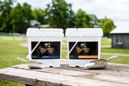
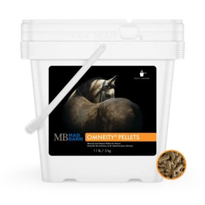
Healthy hoof structure requires adequate amino acid, trace mineral and biotin intake from the diet. Consider feeding our Omneity® mineral and vitamin supplement, which provides a nutritionally effective level of biotin and all other nutrients required to grow out strong, robust hooves.
Omneity® is formulated with 100% organic trace minerals, complete B-vitamin fortification, digestive enzymes, and active yeast cultures. It is a complete mineral and vitamin formula designed to balance the diet of the majority of horses, promote hoof health and enhance digestive health.
Looking for personalized assistance with your horse’s navicular syndrome recovery feeding plan? Submit your horse’s diet for analysis online and Mad Barn’s nutritionists can help for free.
Prognosis
As noted, there is no cure for navicular syndrome. The prognosis depends very much on the individual animal and the level of farrier care. Clinical symptoms may resolve in many cases.
Horses diagnosed with and treated for navicular syndrome may remain serviceably sound for some tasks and not others. For instance, a horse with navicular syndrome may prove fine for riding on the flat but not jumping.
Frequently Asked Questions
Here are some frequently asked questions about navicular syndrome in horses:
Navicular syndrome is a chronic, degenerative cause of heel pain and lameness in horses, usually affecting the front feet. It is also called caudal heel pain or podotrochlear syndrome because it can involve more than just the navicular bone itself. Nearby structures such as the navicular bursa, ligaments, deep digital flexor tendon, and other tissues in the back of the foot may also be involved. [1]
Navicular syndrome is not exactly the same as the older term navicular disease. The newer term is preferred because it reflects that this problem is usually a syndrome involving several structures rather than one isolated disease of the navicular bone. That distinction matters because some horses have heel pain linked more to soft tissues than to obvious navicular bone damage.
The exact cause of navicular syndrome is not fully understood, but repetitive mechanical stress on the heel region is believed to play a major role. Hoof trauma and anything that interferes with blood supply to the navicular area may also contribute. Over time, these stresses can lead to degeneration of tissues and structures in the back of the foot.
Navicular syndrome is more common in heavier, stock-type horses and is seen often in Quarter Horses, Thoroughbreds, and Warmbloods. Horses used for sport or concussion-heavy work such as jumping, reining, and cutting are also at higher risk. Most affected horses are between 7 and 14 years old, and horses kept stalled much of the time may be more vulnerable because reduced movement lowers blood flow to the hoof. [2]
The classic hoof conformation linked to navicular syndrome is a long toe and low heel. Small hooves under a larger-bodied horse can also increase strain on the back of the foot, and upright pasterns may add to the risk. These conformational issues can place more stress on the soft tissues and deep digital flexor tendon in the navicular region.
Horses with navicular syndrome often develop low-grade, chronic lameness that worsens gradually over time. Common signs include a short-strided gait, toe-first landing, difficulty turning, problems going downhill, stumbling, and changes when trotting small circles. Some horses also point a toe at rest, seem stiff through the neck and poll, or become difficult for the farrier to handle.
Navicular syndrome differs from laminitis in the way the horse tries to unload the foot. Horses with navicular syndrome often land toe-first to reduce pressure on the heels, while horses with laminitis usually try to shift weight off the toe. Navicular syndrome also usually does not cause obvious heat, swelling, or a strong digital pulse, so those signs tend to be minimal or subtle.
Navicular syndrome is diagnosed through a lameness examination combined with tests that help confirm the source of heel pain. A veterinarian may watch the horse move on different surfaces, use hoof testers, perform flexion tests, and use a palmar digital nerve block to confirm that pain is coming from the heel region. Imaging is then used to assess the structures involved and the severity of the changes. [3]
Imaging tests used to diagnose navicular syndrome can include radiographs, ultrasound, and magnetic resonance imaging. Radiographs can show bone changes or arthritis, while ultrasound helps evaluate structures such as the deep digital flexor tendon and navicular ligaments. Magnetic resonance imaging is considered the gold standard because it can assess both bone and soft tissues in much greater detail. [8]
Treatment for navicular syndrome usually combines rest, therapeutic shoeing, pain control, and careful long-term management. Horses may need stall rest or confinement to a small paddock at first, especially if tendon or ligament injury is involved. Medications can include non-steroidal anti-inflammatory drugs, corticosteroid injections, or bisphosphonates in horses with bone involvement, but treatment choices depend on which structures are actually affected. [4]
Farrier care is one of the most important parts of navicular syndrome treatment because proper shoeing helps reduce pressure on the navicular region. Therapeutic options may include rolled-toe egg bar shoes to support the heel and encourage easier breakover, or wedge shoes to improve hoof-pastern alignment and reduce tension on the deep digital flexor tendon. Without good hoof balance and consistent follow-up, other treatments are much less likely to succeed. [7]
Shockwave therapy can help some horses with navicular syndrome, but it is not the most effective option for every case. It may help break the pain cycle and improve blood supply, especially when magnetic resonance imaging shows injury to the impar or suspensory ligament. Horses with only one affected front leg may respond better than horses with both front feet involved. [5]
Some horses with navicular syndrome can still be ridden, depending on how severe the condition is and how well they respond to treatment. Many horses can remain usefully sound for lighter work, even if they cannot return to more demanding disciplines such as jumping. The long-term outlook depends on the individual horse, the structures involved, and the quality of hoof and veterinary management.
Summary
Navicular syndrome is a chronic, degenerative cause of heel pain and front‐limb lameness, most common in stock and sport horses, especially Quarter Horses.
- Navicular syndrome involves multiple hoof structures and leads to gait changes such as shortened stride and toe-first landing.
- Risk increases with long toe–low heel hoof shape, small hooves on larger bodies, high-impact work, poor footing, stall confinement, and excess body weight.
- Diagnosis relies on lameness exams with palmar digital nerve blocks and imaging; MRI is the gold standard for assessing both bone and soft-tissue injury.
- Management focuses on corrective farriery (e.g., improving breakover and heel support), workload and footing adjustments, weight control, and targeted therapies such as NSAIDs, corticosteroid or bursal injections, bisphosphonates, and, in refractory cases, shockwave or neurectomy.
- Navicular syndrome is not curable, but early diagnosis and consistent, appropriate care can keep many horses comfortable and usefully ridden for years.
References
- Madsen, L, DVM. Navicular Syndrome in Horses. College of Veterinary Medicine. University of Illinois. Sept. 11, 2020.
- Kentucky Equine Research Staff. Managing Navicular Syndrome in Horses. Kentucky Equine Research. Dec. 13, 2013.
- Pascoe, E. A Pain in the Heels: A New Look at Navicular Disease. Practical Horseman Magazine. Mar. 3, 2014.
- FDA Center for Veterinary Medicine. TILDREN and OSPHOS for Navicular Syndrome in Horses—Information for Equine Veterinarians. FDA. April 21, 2020.
- Brickman-Young, K. Putting shock wave therapy to the test. Western College of Veterinary Medicine. University of Saskatchewan. July 31, 2020.
- Caesars Entertainment Equine Specialty Hospital. Neurectomy-Palmar Digital. College of Veterinary Medicine. Purdue University.
- James K. Belknap. Navicular Disease in Horses - Musculoskeletal System. Merck Veterinary Manual. Oct 2015.
- Widmer, W.R. Use of radiography, computed tomography and magnetic resonance imaging for evaluation of navicular syndrome in the horse. Vet Radiol Ultrasound. 2000. View Summary