
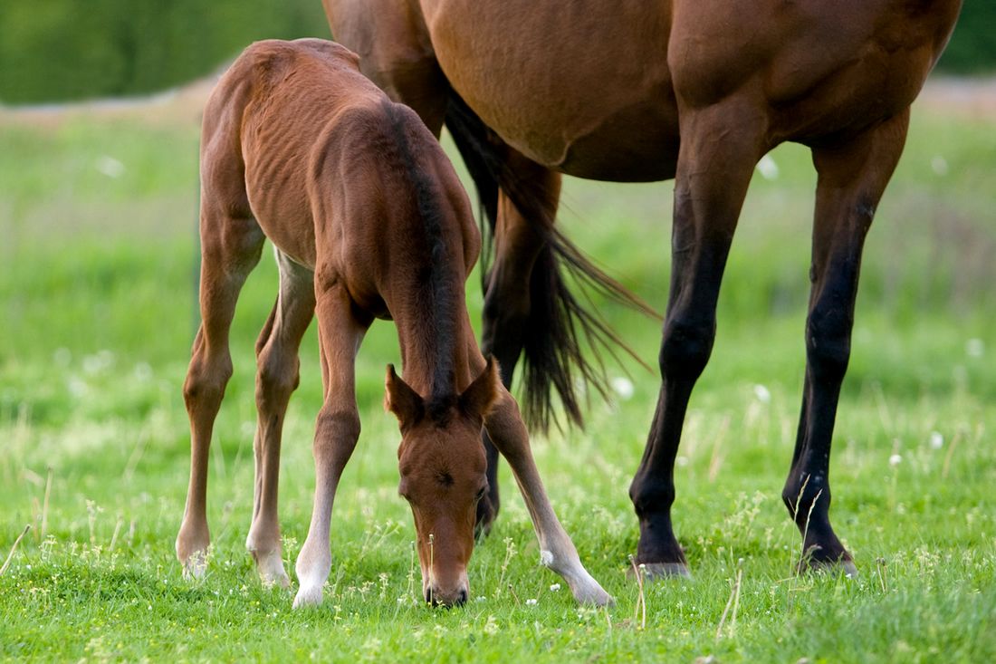
Angular limb deformities (ALDs) and flexural deformities (FDs) frequently affect foals’ legs. When severe, they are deemed clinically significant and require conservative management or veterinary intervention to achieve correction. [1]
ALDs are a deviation from the normal patterns of limb alignment, with lateral or medial angulations usually visible at the fetlock, carpal (knee) or tarsal (hock) level. FDs are joints that cannot be fully extended into a straight standing position.
The causes of ALDs and FDs are multifactorial. Angular limb deformities may develop due to genetics, maternal diseases, premature birth, soft tissue trauma, or nutritional imbalances.
Several treatment options are available once a deformity is recognized, ranging from conservative management by controlled exercise and hoof trimming to more active management such as medical or surgical intervention.
Early detection and intervention are vital to improving the outcome for foals with ALDs and FDs. Mild deformities may be corrected with short-term exercise restriction, and the prognosis for affected horses is generally positive.
Angular Limb Deformities
Angular and flexural deformities in horses can be congenital or acquired.
They are generally classified as congenital when seen in the neonate (newborn foal), having been present from birth. Sometimes the term ‘perinatal’ is used. [2][3]
Acquired ALDs develop over time in response to a primary pathology, which may be developmental or trauma related or due to an unrecognized congenital ALD affecting another joint. [1]
Flexural deformities are often congenital and identified under the misnomer contracted tendons. In the neonatal foal, it is usually observed bilaterally in the forelimbs and is characterized by the inability of the fetlock and carpal joints to straighten. If this remains unresolved, upright hooves may develop.
Types of ALD
ALDs can be categorized into valgus and varus deformities.
- Valgus refers to a lateral or outward deviation of the bone below the joint
- Varus refers to a medial or inward deviation of the bone below the joint. [1][4]
The following deviations are seen in foals:
Carpal valgus
This is a lateral deviation of the third metacarpal (cannon) bone below the knee (carpus), giving a knock-kneed appearance. It is often present in newborn foals, and mild to moderate cases may improve without intervention. [1][4][6]
Carpal varus
This is a medial deviation of the third metacarpal (cannon) bone below the knee, giving a bow legged appearance. It is often associated with lameness on the other forelimb, although it has been seen to develop in weanlings. [1][4][6]
Fetlock valgus
This is a lateral deviation of the phalanges (pastern bones) below the fetlock, giving a toe out appearance. It is common in newborn foals and can be managed conservatively. [1][4][6]
Fetlock varus
This is a medial deviation of the phalanges (pastern bones) below the fetlock, giving a toe in appearance. It is present in newborn foals and is often more serious than fetlock valgus. [1][4][6]
Tarsal valgus
This lateral deviation of the third metatarsal (cannon) bone below the hock gives a cow-hocked appearance in one or both hinds. [1][4][6]
‘Windswept’
Some foals are born windswept, with two limbs deviating in the same direction. This is a valgus in one leg and a varus in the contralateral limb (i.e. affecting both forelimbs and/or both hindlimbs). [1][2][4][5]
It is important to note that different types of ALD may be present at the same time. For example, it is common for a foal presenting a fetlock varus to have a carpal valgus above it. Carpal valgus is often accompanied by rotation at the distal physis of the radius, although the rotation is usually conformational. [1]
Causes of ALD
Asymmetric Growth
The most common cause of ALDs is asymmetric growth in the metaphysis and epiphysis (i.e. growth plate) of the distal radius bone directly above the knee. This can be attributed to a trauma in which a direct impact causes compression and thickening of the physeal plate, with slowed growth.
Any asymmetric loading can lead to asynchronous metaphyseal growth and deviation. Other causes are excessive loading due to lameness in the contralateral forelimb or direct trauma to the joint above the long bone deviation, i.e. fetlock or carpus. [1][4][6]
Incomplete Ossification
Another cause is incomplete ossification of the small carpal and tarsal bones due to premature or dysmature birth. Termed ‘cuboidal’ bones, these begin to ossify late in gestation.
Ossification starts in the centre of the bones and works outward. The carpals are usually slightly rounded at birth, with the final stages of ossification occurring in the first few weeks of life.
If development is delayed when the foal is born, the accessory and fourth carpals are usually the least ossified, allowing the joint to collapse on its lateral aspect. In the hock, the central and third tarsal may be incompletely ossified, contributing to tarsal valgus. [3][6][9][10]
Ligaments
Laxity of the ligaments may also contribute to a carpal valgus.
Periarticular laxity can occur when the foal is born with weak collateral ligaments supporting the joints. A well-conformed neonatal foal will have a mild carpal valgus until its chest expands after a few days.
Weak ligaments can also cause an increase in the angulation, leading to a valgus or varus deformity. This may progress as the carpal or tarsal cuboid bones become malformed. [1]
Endocrine & Nutritional Issues
Chemical toxicity during gestation can also influence the foal’s endocrine system and contribute to ALDs.
Low copper in a pregnant mare’s diet has been shown to lead to higher incidences of ALDs in foals. High or low iodine intake by the mare is another contributing factor. [1]
Some sources have also suggested that malposition in the uterus can lead to ALDs, as well as hormonal or nutritional imbalances. [1]
Heavier birth weight is also associated with ALDs, specifically, carpal valgus and fetlock varus in foals and yearlings. [11]
Diagnosis of ALDs
It is considered normal for a well-conformed neonatal foal to have a mild carpal valgus of 2-5. [2][4][6] In the first month of life, the foal’s chest expands, causing the forelimb angles to straighten and the bones to align. [4][6][10]
Veterinary Exam
A visual examination by a veterinarian is usually sufficient to identify an ALD, by examining the limbs from a perpendicular angle. [2][3][4]
Where rotation accompanies a forelimb ALD, taking a radiograph down the limb to ascertain the relationship between the long bones above and below the joint helps to determine the extent of the deviation attributable to asymmetric epiphyseal growth. [1][3]
Handling and palpation will yield more information. If the ALD is attributable to periarticular laxity, the limb can be straightened manually with the foot lifted. [2][3] If asymmetric epiphyseal growth has occurred, this will not be possible. [1]
In cases when an ALD is attributable to trauma, the foal may be lame, and the affected joint may show signs of effusion. Pain may be present when the joint is palpated. When disproportionate long bone growth is occurring, the foal will display no sign of pain or lameness, although their gait may be affected. [1]
Radiographs
Radiographs provide evidence as to the location, degree of deviation and potentially the cause of an ALD. Images of the growth plate of the radius can confirm asymmetric growth at the metaphyseal plate.
Lines drawn along the axes of the long bones, usually the radius and third metacarpal, show the diagnosed location of the ALD while creating a measurable angle. [1] However, some practitioners maintain that this approach is not always conclusive. [4][12]
Wedging of the epiphysis may also be present on the concave side of deviation, ie. lateral in a carpal valgus presentation. In the fetlocks, medial wedging is often present with a fetlock varus deformity. [1]
Radiographic images of the carpus or tarsus will also reveal excessively rounded bone contours in the case of incomplete ossification, and/or associated malformation of the cuboid bones. [1][4]
What's your top priority with your horse's health?
What's your top priority with your horse's health?
Treatment of ALD
The treatment approach depends on the type of deformity, its location and cause, and the age of the foal. [1]
As carpal valgus can correct naturally in the first weeks of life, most orthopedic veterinarians recommend waiting until the foal is one month old before assessing this ALD. [5]
On the other hand, a fetlock deviation and especially a fetlock varus can be more complex, worsening up to 6 months of age. Due to this and the earlier closure of the distal metacarpal (cannon) physes, these deviations require earlier assessment. [1][2][4][12]
Mild and Moderate ALDs
When a long bone is growing unevenly, the issue will often self-correct. The concave side of the limb will grow faster, and the convex side slower, until the bone evens out.
Foals are usually confined with the mare to ensure that the physis does not become overloaded. If it does, the problem may worsen due to excessive compression. Confinement is continued until the limb has straightened. [1][2][4][12]
Hoof trimming can effectively reduce compressive forces and, combined with conservative exercise, is often sufficient to treat minor to moderate ALDs. [10]
With a fetlock varus, the medial wall of the hoof does not wear as much as the lateral (and vice versa for a fetlock valgus). A small amount of rasping can keep the growing medial wall shorter and in balance with the lateral wall, minimizing asymmetric compressive forces in the limb. [1][2][3][4][6][12]
Extracorporeal Shockwave Therapy (ECSWT) has also been recommended for mild cases. [2]
Severe ALDs
Severe ALDs present in the physis with angles of over 30° of deviation (carpal valgus) or 25° (tarsal valgus) will usually require intervention.
If an ALD is severe at a young age, it may be associated with incomplete ossification of the carpal or tarsal bones. [2][4] The ALD may worsen, and the cuboid bones of the carpus or tarsus may be damaged.
In these cases, surgical intervention is required. Note that rotation of the forelimb associated with ALDs cannot be corrected. [4][6]
HCTP+PS Surgery
Physeal growth is accelerated on the concave side of the bone through hemicircumferential transection of the periosteum and periosteal stripping (HCTP+PS).
There is discussion about the benefits of HCTP + PS compared with frequent trimming, although many veterinarians support its use for mild to moderate cases where conservative methods have failed.
It is easy to perform, inexpensive, and has very few complications. Athletic potential is usually realised in a case involving a single surgery, although it is impaired following surgery at multiple sites. [1][4][6][12]
Transphyseal Bridging
Growth is slowed, through transphyseal bridging (TPB) surgery on the convex side of the deformity. This works through a process of static compression.
Physeal growth is restricted by bridges through the physis made of staples, screws or wire. Alternatively, a small metal plate may be screwed across the physis.
Endochondral ossification is slowed on the convex side of the bone, while growth continues at the same rate on the concave side of the bone. Eventually, the limb becomes straight.
Results are generally positive, although this surgery is more complex to perform. A second intervention is needed to remove the implants, and there is always the risk of over-correction if this is not done at the correct time. [1][4][6][12]
In some severe cases, growth acceleration and reduction techniques are both used to achieve a faster, more complete correction. [12]
Rarely, osteotomy techniques are used, with sections of physeal bone removed in ‘closing wedge’ and ‘step’ procedures. [2][12]
Timescales for ALD surgery
The order in which growth plates adjacent to different joints close and the age of the foal determine the timing of surgery.
ALDs of the fetlock or phalanges (pastern and hoof joints) should generally be treated surgically within 45 – 60 days of age, with HCTP+PS performed by 1 – 2 months and TPB by 3-4 months. Minor ALDs at the fetlock level may be treated at up to 6 months of age. [6]
With ALDs involving the knees and hocks, (i.e. those of the distal radial physis in the forelimb and distal tibial physis in the hindlimb) surgery is usually arranged at 4 – 5 months. TPB is most effective for tarsal valgus at 60 days or earlier. It is often performed in foals above 4-5 months with carpal valgus greater than 15°. [2]
Despite these guidelines, many veterinarians leave surgery until later with all but severe carpal valgus deformities. In mild to moderate cases, conservative management and natural correction is usually a first step. [1][8]
Some believe treatment is effective at any point before the closure of the distal radial physis (i.e. before 60 weeks of age). [10]
Prognosis for ALD
Foals with mild to moderate valgus ALDs affecting the fetlocks, carpals or tarsals are likely to recover well with appropriate management, with many achieving full athletic soundness. [1][6]
Varus deformities are more difficult to correct, and this may lead to pastern and fetlock lameness. [1][10] This has been found to negatively affect athletic outcomes in racehorses.
On the other hand, a mild carpal valgus may be protective in athletic horses, reducing the incidence of interference injuries from the hind hooves in a gallop. [3][13]
Moderate ALDs can contribute to wedging of the carpal and tarsal cuboid bones in the early months.
Flexural Deformities (FD)
A flexural deformity in foals involves the inability of the fetlocks and knees to fully straighten, leading to the development of upright hooves. It usually involves the knee or the knee and fetlock, but it is also occasionally seen in the pasterns.
The cause of FDs rests either in the muscles and tendons above or other abnormality. More than one structure may be involved, including the ligaments around the affected joints. [4][7]
FD is either congenital or develops during a foal’s growth period. It can be related to a traumatic injury to one limb, which causes the youngster to flex and reduce weight bearing.
An upright hoof develops as a result of being partially loaded for a prolonged period of time. [1] As an acquired deformity, it is also often seen in weanlings during a rapid growth period. [7][8]
Some researchers also categorize congenital digital hyperextension, popularly termed lax tendons or dropped fetlocks, as a flexural deformity. [8] This usually resolves after being managed conservatively and is not within the scope of this article.

Causes of FD
Contraction of the tendons is not a cause of flexural deformities.
Congenital FDs
When congenital, FDs may be influenced by uterine mal-positioning, growth restriction, genetic or nutritional factors.
Rarely, a condition called arthrogryposis may be a congenital cause. [1] Where the carpals are involved, it may be part of a larger picture of foal contraction or dystocia. [4]
Acquired FDs
Acquired FDs may be accompanied by rupture of the common digital extensor tendons. This can occur either prior to the development of an FD, or as a result of it. It is usually detectable in bilateral swellings on the outside of both knees. [1]
Unilateral FD can develop as a result of traumatic injury to one limb. The unloaded limb may develop an upright hoof through flexure of the phalangeal (pastern and hoof). The foot may knuckle forwards and over if the fetlock is also involved.
Pathological Factors
Primary pathological factors include physitis of the distal radius and Osteochondritis dissecans (OCD) in upper joints. Trauma to the flexor tendons may also contribute to FD, due to the restriction caused by scar tissue.
Over-nutrition of the mare, foal or both during the suckling and growth period may result in rapid foal growth.
It is believed by some that there are discrepancies in growth rates between bone and ligaments or between the strength of flexor and extensor muscles, although this is debatable. [1][4] Others maintain that there is strong evidence for physeal pain being a cause. [4][7]
Diagnosis of FD
An upright foot is usually visually evident. Heels usually remain on the ground but, in severe cases, may be raised with the foal walking on the toe only. Lameness is usually absent.
The degree of the FD is determined by the angle of the dorsal (toe) wall of the hoof: [1]
- Stage I is defined as prior to the dorsal wall becoming vertical
- Stage II is defined as beyond vertical and requires a more aggressive treatment
Grading Clubfeet
An additional system for grading clubfeet exists:
- A Grade 1 clubfoot has a hoof angle 3–5° greater than the contralateral foot. The hoof-pastern axis remains aligned.
- A Grade 2 clubfoot has a hoof angle 5–8° greater than the contralateral foot. The hoof-pastern axis is steep and slightly broken forward.
- A Grade 3 clubfoot has a broken-forward hoof-pastern axis. The dorsal hoof wall may be dished.
- A Grade 4 clubfoot has a hoof angle of 80 and a severely broken-forward hoof-pastern axis. [7]
Examination Findings
Deformity at the fetlock level (metacarpophalangeal joint) may involve the deep digital flexor tendon, the superficial digital flexor tendon, and the suspensory ligament.
Palpation will determine which flexor tendon is the tightest. This is subjective, but may determine which tendon to treat.
This may be confirmed through radiographs. These may reveal osteolysis and remodelling of the third phalanx, or pedal, bone. [1][7]
Deformity at pastern level (the distal interphalangeal joints, or DIP) usually involves only the deep digital flexor tendon. [1]
Treatment of FD
Minor congenital FD may self-resolve without treatment. Some FDs may improve simply with bandaging, splinting, or corrective shoeing. If the extensor tendons are ruptured, these may heal with confinement alone, with splinting to prevent the fetlock from knuckling. [1]
More severe FDs may require medical or surgical management soon after diagnosis, as the foal may have difficulty standing, in which case the deformity will worsen.
Veterinary treatment usually commences in the young foal with NSAIDs and intravenous oxytetracycline, along with bandaging and splinting. Intravenous oxytetracycline helps relax tendon tissue, and may resolve minor cases of flexural deformity. Oxytetracycline may be administered 2 to 3 times, but there is a risk of renal issues with increased doses. [4][7]
Tenotomy & Transection
If the above fails, then surgical treatment in the form of a tenotomy (detaching tendon from bone) or transection (cutting across tendon) may be necessary.
- Tenotomies of the flexor tendon, inferior check ligament (for flexure of the fetlock) or superior check ligament (for flexure of the knee) may be used but are not usually necessary.
- Tenotomy of the deep digital flexor tendon is usually reserved for horses over 12 months of age, with severe flexure at fetlock level.
- Transection of the superior check ligament may help foals that are chronically over at the knee, as it extends the joint.
- Transection of the palmar carpal ligament at the back of the knee may also help foals with serious flexure at the knees.
- Tenotomy of tendons above the knee (ulnaris lateralis and flexor carpi ulnaris) can help moderate flexure at the knee. [4][7]
The surgery selected for fetlock FDs depends on subjective assessment of the soft tissue structures, as well as angles.
- Mild FDs with angles less than 180° are usually treated with an inferior or superior check ligament desmotomy (surgical cut to lengthening of the tissue).
- Moderate FDs with angles of 180° are upright and are treated with an inferior or superior check ligament desmotomy, with splinting during recovery.
- Severe FDs with angles of greater than 180° and that are knuckling forward are treated with an inferior check ligament desmotomy as well as either a superior check ligament desmotomy or superficial digital flexor tendon tenotomy, with splinting after surgery. [1]
Prognosis for FD
Foals with mild flexural deformities typically have a good prognosis with appropriate medical management. Similarly, foals treated before the age of 6 to 8 months with inferior check ligament desmotomy have a positive prognosis for normal hoof conformation and athletic soundness.
Transection of the inferior check ligament does not have significant longer-term effects on locomotion, joint angles, or other tendons and ligaments.
Correct management and rehabilitative shoeing or trimming of the heel with hoof extensions supports recovery. [1][2]
Foals with moderate severe flexural deformities have a poor prognosis for future athletic performance. Some horses may be comfortable for light riding. Individual assessment by a veterinarian is necessary to determine athletic potential of these horses once they are fully grown.
Frequently Asked Questions
Here are some frequently asked questions about angular and flexural limb deformities in foals:
Angular limb deformities are visible outward or inward deviations of a foal’s legs, most often seen at the knees, fetlocks, or hocks. Flexural deformities involve joints that cannot fully straighten, giving the appearance of contracted tendons or upright pasterns. Both conditions can range from mild to severe and may be present at birth or develop as the foal grows. Early recognition and monitoring are essential to prevent long-term effects on soundness.
ALDs can develop for many reasons, but they usually stem from uneven growth at the growth plates of the long bones. Premature birth, incomplete bone ossification, trauma, or uneven weight-bearing on one limb can all contribute. Nutritional imbalances during gestation, such as low copper or inappropriate iodine levels, have also been linked to higher risk. In some cases, foals may inherit a predisposition, or the condition can result from restricted positioning within the uterus.
Flexural deformities can appear at birth or develop later in the growing foal. Congenital cases often arise from limited space in the uterus, genetic factors, or developmental issues that affect the muscles and tendons. Acquired deformities, on the other hand, may occur due to pain, injury, or rapid growth that causes the tendons to tighten faster than the bones lengthen. Overnutrition or imbalanced diets during growth can also increase the likelihood of developing these deformities.
Preventing limb deformities begins with balanced nutrition for the mare during pregnancy and the foal after birth. Foals should receive adequate turnout and controlled exercise to strengthen joints and muscles without overloading developing limbs. Regular farrier care helps maintain proper hoof balance, reducing uneven stress on growing legs. Most importantly, any signs of limb deviation or stiffness should be assessed early by a veterinarian, as prompt management offers the best chance of full correction.
Summary
Angular limb deformities (ALDs) and flexural deformities (FDs) are common leg abnormalities in foals that affect limb alignment and joint flexibility. Early diagnosis and timely intervention are key to successful correction and future soundness.
- ALDs involve outward (valgus) or inward (varus) limb deviations, often seen in knees, fetlocks, or hocks
- FDs restrict joint extension, causing upright or knuckled-over hooves in foals
- Contributing factors include genetics, trauma, premature birth, and nutritional imbalances
- Mild deformities may resolve with rest, trimming, or controlled exercise
- Severe cases may need surgical correction such as periosteal stripping or transphyseal bridging
- Balanced nutrition and proper mare diet during gestation help reduce deformity risk
- Most foals with early treatment achieve normal limb alignment and athletic potential
References
- Baxter GM. Angular Limb Deformities (ALDs) In: Baxter GM, ed. Adams and Stashak’s Lameness in Horses. 6th ed. Sussex, UK: Wiley-Blackwell, 2011;1138-1144.
- Smith L. Treatment of angular limb deformities in foals. In Practice. 2010. 32.
- Witte S, Hunt R. A review of angular limb deformities. Equine Veterinary Education. 2009. 21:378-387.
- Greet TRC, CertEO. Managing Flexural and Angular Limb Deformities. The Newmarket Perspective. 1997.
- McIlwraith C, Anderson, TM, Sanschi, EM. Conformation and musculoskeletal problems in the racehorse. Clinical Techniques in Equine Practice. 2003. 2:339-347.
- Peat FJ, Kawcak CE. Musculoskeletal Pathology. Veterinary Clinics of North America: Equine Practice. 2015. 31:407-424. View Summary
- O'Grady SE. Flexural deformities of the distal interphalangeal joint (clubfeet). Equine Veterinary Education. 2012. 24:260-268.
- Robert C, Valette JP, Denoix JM. Longitudinal development of equine forelimb conformation from birth to weaning in three different horse breeds. The Veterinary Journal. 2013. 198, Supplement 1:e75-e80. View Summary
- Clothier J. Beyond the Miracle Foal: A Study into the Persistent Effects of Gestational Immaturity in Horses (Doctoral Thesis). Armidale, Australia: University of New England. 2019.
- Engiles JB, H. Stewart, J. Janes, L. A. Kennedy. A diagnostic pathologist’s guide to carpal disease in racehorses. Journal of Veterinary Diagnostic Investigation. 2017. 29:414-430. View Summary
- Santschi EM, Leibsle SR, Morehead JP, et al. Carpal and fetlock conformation of the juvenile Thoroughbred from birth to yearling auction age. Equine Veterinary Journal. 2006. 38:604-609. View Summary
- Auer JA, von Rechenberg B. Treatment of Angular Limb Deformities in Foals. Clinical Techniques in Equine Practice. 2006. 5:270-281.
- Love S, Wyse CA, Stirk AJ, et al. Prevalence, heritability and significance of musculoskeletal conformational traits in Thoroughbred yearlings. Equine Veterinary Journal. 2006 38:597-603. View Summary
- Anderson TM, C.W. McIlwraith. Longitudinal development of equine conformation from weanling to age 3 years in the Thoroughbred. Equine Veterinary Journal. 2004. 36:563-570. View Summary


