
Equine asthma (EA) is the collective term for chronic respiratory signs in horses that range in severity from mild to severe.
These conditions were previously known as inflammatory airway disease (IAD) or recurrent airway obstruction (RAO), respectively.
Equine asthma is characterized by inflammation and mucous production in the lungs, which leads to lower airway obstruction. The horse may cough, and breathing may or may not be laboured.
Asthma is most commonly observed in horses stabled over winter months that have become hypersensitive to dust, airborne allergens, mold spores, or mites in stable bedding or stored hay. [1][2]
Unstabled horses may also react to pollens when at pasture in summer, with the condition known as Equine Pasture Asthma (EPA). [3]
The formerly used terms Chronic Obstructive Pulmonary Disease (COPD) and Summer Pasture Associated Obstructive Pulmonary Disease (SPAOPD) are now obsolete. [2][3][4]
Other terms that have been used interchangeably in the past include equine emphysema and chronic bronchiolitis. [5] These conditions are also now grouped under equine asthma. [4][5]

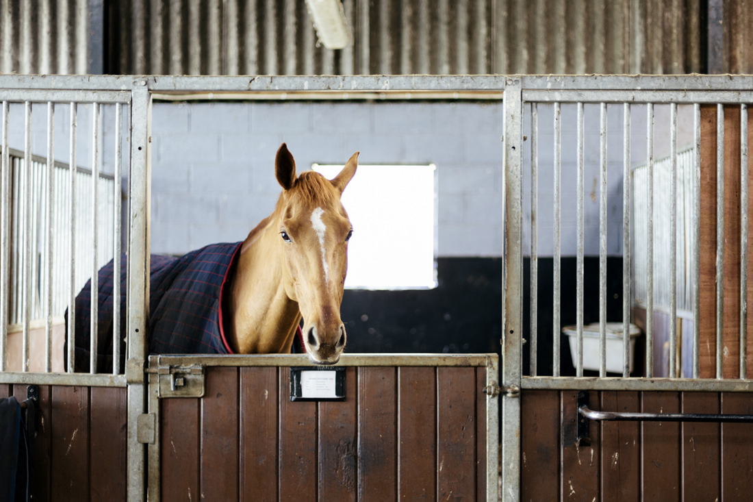
Equine Asthma
Like some forms of human asthma, equine asthma has an immunological basis. The disease is strongly linked to environmental dust, pollen, and in some cases to preceding bacterial and viral infections.
The exact mechanisms of this immunological response are not yet fully understood, nor is it known why only some horses are affected. [4] Individuals of all ages, breeds and both sexes are affected, although it does appear more frequently in older horses. [6][7]
In horses over 20 years, a declining immune function may be a factor, with a tendency towards the exaggerated inflammatory responses that underpin this disease. [7]
Younger horses may have been affected by early immune events, with the memory component of the adaptive immune system being repeatedly triggered by the same antigen. [7] As clinical cases are usually more advanced or late stage in older animals, it is difficult to identify the earlier disease processes. [3]
Additionally, a genetic factor has been identified in asthmatic horses. The presence of a major gene in families demonstrates that a predisposition to EA can be inherited. [6]
As yet, there is no cure for equine asthma, but in most cases, a combination of treatment and environmental management can bring about reversal through clinical improvements.
Types of Equine Asthma
Asthma in horses is usually classified in one of two ways, based on the degree of hypersensitivity to allergens affecting the lungs:
- Mild to moderate EA: mainly subclinical, making diagnosis challenging
- Severe EA: has clear clinical signs and is often chronic, recurring seasonally and worsening if inadequately treated
Environmental Conditions
Equine asthma is commonly found in stabled horses exposed to dust and molds from poor-quality hay and bedding for long periods of time in winter.
Over 50 different bacterial endotoxins, mold species, microbial toxins, storage mites, plant particles, other microbes, and inorganic dust have been identified in stable dust. [2] Affected horses are often aged over 7 years. [4]
Horses at pasture in summer may present mild to moderate signs of equine pasture asthma in response to pollen, tropical grasses, fungal spores, dust from harvesting, or crop burning. [4]
Both mild and severe asthma are strongly linked to environmental dust, and in some cases to earlier bacterial and viral infections such as: [4]
- Equine influenza virus (EIV)
- Equine herpesviruses (EHV)
- Equine rhinitis viruses (ERVs)
- Streptococcus equi subspecies zooepidemicus
- Actinobacillus
- Pasteurella spp
Occasionally, horses may present severe forms of both EA and EPA. [4]
Pathophysiology
As a condition of the lower respiratory airway, equine asthma has a similar pathophysiological progression to its human counterpart. The exact processes involved are unclear, although it is thought to be a multifactorial process.
In non-asthmatic horses, dust particles are expelled from the lungs towards the nasopharynx. If dust particles remain in the lung, they are effectively dealt with by the alveolar macrophages.
These are white blood cells, also known as ‘dust cells’, which form the first line of defence against pathogens in the lung. [8]
Allergic Hypersensitivity
In asthmatic horses, an elevated inflammatory response occurs, and immunoglobulin E (IgE) antibodies are produced by plasma cells. T-cells (a type of white blood cell) are also recruited into the lungs. [3]
With this allergic response, several things happen.
- The tissues become inflamed. This is due to an increase in neutrophils (a type of white blood cell), which fight infection.
- The presence of neutrophils also drives mucus production. This is thick and contributes to airway restriction and blockage. [4]
- In severe cases, inflammation and mucus production fill the airways, resulting in partial airway obstruction.
- The smooth muscles that line the airways constrict and tighten (bronchospasm), causing an increase in muscle mass.
- With ongoing bronchospasm, the muscles become hyperplasic, ie. spasmed, hard and enlarged. Some horses have 300% more peripheral airway smooth muscle than before, and 50% more in central airways. [3]
- Fibrosis (scar tissue formation after inflammation) restricts the peripheral airways even more. Over time, permanent tissue remodelling may occur. [4]
- Damage to the alveoli (air sacs in the lungs) can occur, causing the inner walls of the sacs to rupture. This leads to emphysema, with permanent shortness of breath. [4]
What's your top priority with your horse's health?
What's your top priority with your horse's health?
Signs & Symptoms
Equine asthma may have a sudden or gradual onset. It often coincides with a management change, such as when the horse is brought in from the pasture to the stable. Sometimes more than one horse in a stable may be affected.
Equine pasture asthma can commence with the summer months and a rise in pollen. [9]
Mild Asthma
Mild asthma in horses can be subclinical and hard to detect, with possible signs including:
- Exercise intolerance at higher gaits
- Reduced performance at higher levels
Moderate Asthma
Moderate equine asthma presents additional signs to the above, including:
- Mild nasal discharge
- Labored breathing after exercise
- Coughing during or after exercise (not all horses present this symptom)
The condition can be progressive if untreated and the horse is continually exposed to the allergens.
Severe Asthma
Severe asthma in horses is characterized by the following commonly reported signs:
- Labored breathing at rest (dyspnea) with abdominal lifting (previously known as heaves)
- Mucoid nasal discharge
Clinical signs of severe EA detectable through veterinary diagnostics include: [4]
- Increased blood pressure
- Abnormal respiration sounds, ie. crackles, wheezes, etc., on either one side or both, audible through auscultation (listening with a stethoscope)
- Increased diameter of the pulmonary artery, visible on ultrasound
- Increased right ventricular wall thickness, visible on cardiac ultrasound
Diagnosis
Differential Diagnosis
Other conditions affecting the lungs can have similar symptoms to equine asthma, including: [9]
- Exercise induced pulmonary hemorrhage
- Lungworm
- Neoplasia
- Pleuritis
- Pneumonia
- Tracheal deformity
- Foreign body
- Tracheobronchial tuberculosis
Case History
A tentative diagnosis can be made based on the owner-provided history. This may immediately reveal that equine asthma episodes correlate to seasonal triggers.
This is confirmed if the horse improves within hours or days of being isolated from the triggering stable or pasture.
Case history may also reveal other risk factors, including whether the horse’s sire or dam is affected by equine asthma, what forage the horse is fed, and how the feed is delivered. [4]
A history of febrile (fever-causing) illness is also relevant, as well as any recent viral infection. [9]
While case history can be a compelling element for diagnosing severe EA, more diagnostics are typically needed for mild or subclinical EA. [4]
Auscultation
Listening to the lungs following light exercise may reveal crackling lung sounds in mild EA cases. A simple rebreathing bag may yield the same results. [9] Further tests can then take place.
Tracheal Endoscopy & Tracheal Wash
Practicing veterinarians commonly prefer to use the combination of tracheal endoscopy and tracheal wash. [4]
Tracheal endoscopy enables the visual detection of mucus in the lower airways. It offers a reliable diagnosis for mild to moderate asthma of fewer than 3 weeks duration, as well as severe asthma. [4]
A tracheal wash involves collecting a fluid sample from the trachea for cytological examination. As well as enabling pathological confirmation in the laboratory, it allows sequential sampling to monitor disease progression and remission. [4]
Endobronchial Biopsy
Endobronchial biopsies are easy to obtain, and when processed for standard histology, they allow the assessment of airway epithelium, lamina propria and smooth muscle.
However, they do not permit a precise quantification of the deepest bronchial structures (smooth muscle, cartilage and peribronchial tissues). [5]
A limitation is that biopsies show no clear difference between severe EA cases in remission and unaffected horses. [4]

Endobronchial Ultrasound (EBUS)
This is a technique for imaging the bronchial wall using a probe to evaluate the inflammation and remodelling affecting all the airway structures. [5]
Lung Function Testing
Some specialist equine clinics offer lung function testing, which is the ultimate tool for assessing and tracking asthma-affected respiratory function in humans.
Lung function tests can detect severe EA, but are less useful for mild cases in which the baseline lung function is often unaffected. [4]
Bronchoalveolar Lavage (BAL)
This method of examining airway secretions is effective for diagnosing mild, moderate and severe EA.
Saline is infused into the lungs and collected to measure neutrophils, mast cells and eosinophils. Affected horses will have an increase in neutrophil and lymphocyte percentages. [3]
However, there is a lack of established criteria for diagnosing the disease and no set testing protocols for bronchoalveolar lavage. [10]
Other Tests
Several commercial serological tests for identifying asthma in horses involve measuring antibody levels, such as serum-free immunoglobulin E.
However, there is debate over the diagnostic value of these tests, due to a lack of consistent trial results. [2]
There are also several commercially available bronchial provocation tests, which aim to identify the allergens involved in triggering the asthma response.
In positive cases, these tests cause airway obstruction within hours of administration, but their reliability has also been questioned. [2]
Treatment
Once a diagnosis has been achieved, a treatment plan is prepared, based on the severity and type of equine asthma. Treatment goals involve reducing the challenge to the immune system and suppressing excessive allergic responses.
Medication is usually a combination of bronchodilators, corticosteroids, antibiotics, and, if necessary, antihistamines. [11]
Bronchodilator drugs bring short-term relief from episodes of acute respiratory distress and bronchospasm. As in humans, a Ventopulmin® bronchodilator should provide rapid relief within 30 minutes. [5]
Corticosteroids
Corticosteroids address the inflammation that causes the narrowing of the airways. [12] These can be administered through inhalation, orally, or through intramuscular injection.
Injected or ingested (systemic) corticosteroids are often more effective in bringing rapid relief. [11] Both injected and inhaled corticosteroids have a limited ongoing effect after the treatment phase ends.
- Dexamethasone is highly potent, typically relieving severe symptoms within 3 days.
- Prednisolone is less effective than Dexamethasone. [11]
- A single dose of Triamcinolone acetonide is both potent and long-acting for approximately 4 weeks. [11]
- Methylprednisolone acetate has a milder, transient effect. [11]
- Isoflupredone acetate is as effective as Dexamethasone in improving lung function but is associated with a risk of hypokalemia. [11]
Environmental Management
Appropriate environmental management is required to achieve full remission from equine asthma and prevent disease progression.
Medication addresses the signs of asthma and speeds recovery, but reducing exposure to allergens is essential. [9] Studies show that cytology remains abnormal until management changes. [5]
Managing a horse with asthma usually means removing the horse from the stable environment where the disease becomes apparent or removing them from pasture when pollen count is high.
In severe cases, the horse may even be isolated in a climate-controlled environment. Managing environmental factors can present considerable difficulties to horse owners. [4]
With proper management, many non-severe cases recover and remain asymptomatic.
Management Changes
For stabled horses, management changes should also include some or all of the following measures: [9]
- Ensuring fresh air is available, with good ventilation for clearance of dust particles.
- Removing horses from the stables during mucking out, when highest dust levels occur.
- Using alternatives to straw for bedding, including shavings, sawdust, shredded paper, etc.
- Turning horses out to pasture if possible.
- Eliminating hay if possible, replacing with silage or haylage (always check for damaged plastic packaging), or cubed lucerne.
- If hay has to be fed, purchasing bales with a low moisture content, preferably that has been sun dried for 2-3 days before baling.
- Storing hay away from affected horses, with spaces between bales for aeration.
- Soaking hay for 15 minutes and feeding it with 30 minutes of soaking.
- Moving manure away from stable vicinity every 3-4 days.
Prognosis
Equine asthma is not curable, but full remission can certainly be achieved. Many horses can return to normal lung function, even when there has been remodelling in the airways.
However, emphysema or shortness of breath may remain present in some severely asthmatic horses. [13] Lung elasticity can be permanently affected by tissue remodelling.
This causes stiffness in the peripheral airways, leading to ongoing respiratory issues and exercise intolerance, even when the horse is isolated from seasonal triggers. [4] In such cases, athletic careers are compromised or curtailed.
Asthma is rarely fatal in horses, unless complications develop. [4] An example of a complication is cor pulmonale, or right side heart failure, which can occur as a result of ongoing hypertension (high blood pressure) within the lungs due to airway restriction and lowered oxygen levels. [14]
Severe equine asthma can compromise quality of life, especially if a treated horse continues to experience respiratory distress while at rest and cannot exert itself athletically. [9]
In these cases, it is common for owners to euthanize severely affected horses due to a combination of factors: the expenses of ongoing therapy, the difficulty in maintaining a dust-free or low-dust environment, and significant quality-of-life concerns. [15]
If your horse has been diagnosed with equine asthma, work with a nutritionist to formulate a low-dust diet that supports respiratory health. Submit your horse’s information online for a free consultation.
Frequently Asked Questions
Here are some frequently asked questions about equine asthma in horses:
Recognizing if a horse has asthma often begins with noticing the shift in its breathing or comfort during normal activity. Many horses show coughing, reduced tolerance for work, or mild nasal discharge as inflammation and mucus develop in the lower airways. Some progress to labored breathing after exercise or even at rest. Seasonal changes or a recent move into a dusty barn can make these signs more obvious.
Environmental features that result in asthma flare-ups usually involve airborne irritants building up around the horse. Dust from hay, bedding, or enclosed winter stabling introduces molds, mites, and plant particles that trigger airway inflammation. Pasture conditions can create similar problems when pollen, grasses, fungal spores, or harvest dust increase during summer. Continued exposure tends to intensify the allergic response.
Full recovery from asthma depends on reducing the immune challenge that drives airway inflammation. Many horses improve dramatically with medication and strict management that limits dust and seasonal allergens, sometimes returning to normal lung function. Long-standing or severe disease may leave residual airway stiffness or emphysema, which narrows the chance of complete reversal. Careful management still keeps most non-severe cases comfortable and functional.
Summary
Equine asthma occurs when a horse’s lower airways react to inhaled particles or allergens, creating inflammation that can make breathing harder and limit performance. Symptoms may appear during certain seasons, after changes in management, or gradually worsen if the environment continues to challenge the lungs.
- Susceptibility varies, but many cases begin with sensitivity to dust, molds, pollens, or other airborne irritants influenced by both immune and genetic factors
- Inflammatory reactions inside the lungs cause mucus buildup, narrowing of the airways, and tightening of the surrounding muscles that restrict airflow
- Veterinarians may rely on endoscopy, fluid sampling, rebreathing evaluations, and specialized imaging to determine the extent and type of airway involvement
- Treatment usually pairs medications that reduce inflammation or open the airways with management steps that limit exposure to problem particles
- Most horses improve with consistent care, though severe or long-standing disease can leave structural airway changes that affect long-term performance
References
- Managing COPD. Journal of equine veterinary science. 1998;18(10):650-650.
- Klier, J. et al. Comparison of Four Different Allergy Tests in Equine Asthma Affected Horses and Allergen Inhalation Provocation Test. Journal of equine veterinary science. 2021;102:103433-103433. View Summary
- Bowles, K.S. et al. A novel model for equine recurrent airway obstruction. Veterinary immunology and immunopathology. 2002;87(3):385-389. View Summary
- Couetil, L. et al. Equine Asthma: Current Understanding and Future Directions. Frontiers in Veterinary Science. 2020-July-30 2020;7.
- Bullone, M. and Lavoie, J-P. Equine asthma diagnosis: Beyond bronchoalveolar lavage cytology. Equine Veterinary Journal. 2017;49(3):263. View Summary
- Gerber, V. et al. Mixed Inheritance of Equine Recurrent Airway Obstruction. Journal of veterinary internal medicine. 2009;23(3):626-630. View Summary
- Hansen, S. et al. A review of the equine age-related changes in the immune system: Comparisons between human and equine aging, with focus on lung-specific immune-aging. Ageing research reviews. 2014;20:11-23.View Summary
- Hu G. and Christman J.W. Editorial: Alveolar Macrophages in Lung Inflammation and Resolution. Frontiers in Immunology. 2019-September-24 2019;10.
- Clarke A. Lower Respiratory Tract Diseases: COPD. In: Robinson N, ed. Current Therapy in Equine Medicine: WB Saunders Company. 1992.
- Couëtil, L. et al. Inflammatory Airway Disease of Horses--Revised Consensus Statement. J Vet Intern Med. 2016. View Summary
- Mainguy-Seers, S. and Lavoie, J-P. Glucocorticoid treatment in horses with asthma: A narrative review. J Vet Intern Med. 2021. View Summary
- Rossdale, P. and Ricketts, S.W. Equine Stud Farm Medicine. Philadelphia: Lea & Febiger; 1980.
- Derksen, F.J. et al. Comparison of transtracheal aspirate and bronchoalveolar lavage cytology in 50 horses with chronic lung disease. Equine Vet J. 1989. View Summary
- Dixon, P.M. and McGorum, B.C. Equine pulmonary disease: a case control study of 300 referred cases. Part 2: Details of animals and of historical and clinical findings. Equine Vet J. 1995. View Summary
- Couëtil, L. Analysis of risk factors for recurrent airway obstruction in North American horses: 1,444 cases (1990-1999). J Am Vet Med Assoc. 2003. View Summary


