English
Joint disease and injury are common causes of lameness and poor performance in horses, particularly in athletic or young, growing animals. Conditions such as osteochondritis dissecans (OCD), synovitis, and traumatic fractures of joint surfaces can significantly affect a horse’s comfort, movement, and long-term soundness.
Arthroscopy has become the gold standard for addressing these issues because it allows the surgeon to assess the joint directly, rather than relying solely on imaging techniques such as radiographs or ultrasound. This procedure involves inserting a small camera and specialized instruments through tiny incisions to visualize, diagnose, and treat problems inside the joint.
Arthroscopy offers many advantages over traditional surgical techniques. Small incisions associated with the procedure reduce soft tissue trauma, postoperative pain, and infection risk. Arthroscopy also carries faster healing times with minimal scarring compared to more invasive methods.
As equine athletes continue to push the boundaries of performance, maintaining joint health has become a top priority in both prevention and treatment. Arthroscopy represents a key advancement in this effort, combining precision, safety, and effectiveness to support better long-term outcomes for horses of all disciplines.
Arthroscopy for Horses
Arthroscopy is a minimally invasive surgical procedure that allows direct visualization of the inside of a horse’s joint using a small camera which transmits real-time images to a monitor. By making multiple small incisions, specialized instruments can be inserted at the joint site to remove loose fragments, smooth damaged cartilage, or perform other delicate procedures.
Arthroscopy can be used for diagnosis and treatment of joint problems in horses. As a diagnostic tool, arthroscopy allows the veterinarian to evaluate the cartilage, bone, and soft tissues making up a joint. [1]
Arthroscopy is used to diagnose numerous conditions, including: [1]
- Osteoarthritis
- Osteochondritis dissecans
- Synovitis
- Ligament injuries
- Cartilage fragments
In most cases, these conditions are strongly suspected based on the results of other diagnostic tests, including physical examination, X-rays, ultrasound, MRI, or CT. [2] Arthroscopy allows for definitive diagnosis of these conditions in cases where the other diagnostic modalities are inconclusive. [2]
During treatment, surgeons can remove damaged tissue and fragments of cartilage or bone to restore normal joint function. Arthroscopy is most commonly used to treat osteochondral fragments from osteochondritis dissecans, but the other conditions listed above can also benefit from arthroscopy. [1]
What's your top priority with your horse's health?

What's your top priority with your horse's health?
Equipment
The main piece of equipment for arthroscopy is an endoscope, a rigid tube with a camera or optical unit on the end that the surgeon inserts into the joint. [1] Most endoscopes used for arthroscopy in horses are under 4 mm in diameter. [1]
The endoscope transmits the image to an eyepiece or external monitor for the surgeon to view. [3] The endoscopy unit also has a light source to illuminate the tissue for visualization. [1]
The rigid tube that makes up the endoscope is hollow, allowing the surgeon to introduce fluid or gas into the joint capsule. [1] This helps clean tissue surfaces, remove bleeding, and pressurize the joint capsule to maximize visibility. [1]
If using arthroscopy for treatment, the surgeon introduces surgical tools into a second incision in the joint space. [1] Instruments may include forceps, bone curettes, blades, or probes. [1]
Procedure
Most surgeons perform arthroscopy while the horse is under general anesthesia. After anesthetizing the horse and moving them into the surgical suite, they begin preparing the skin for the procedure.
Depending on the location of the surgery and surgeon preference, skin preparation may include clipping or shaving the hair. [2] The veterinary team scrubs the skin with antiseptics like povidone iodine or chlorhexidine to remove any bacterial contamination on the skin surface. [2]
Once the skin is clean, the veterinary team places waterproof drapes around the surgical site. These drapes help prevent contamination of the surgical site during the procedure. [2]
The surgeon makes an initial incision near the joint and inserts the arthroscope. They use fluid or gas to inflate the joint capsule, making it easier to maneuver instruments within the joint space. [2]
Depending on visibility, the surgeon may have to flush the joint by introducing fluid and allowing it to drain out of a second incision or a needle placed through the skin. [2] This process removes any material that may prevent assessment of the joint tissues.
Once the joint space is clean, the surgeon begins evaluating the joint for any changes that may cause lameness. After the evaluation is complete, the surgeon may proceed with treatment depending on their findings.
Treatments may include: [2]
- Removal of bone or cartilage fragments
- Debriding (removing) tissue to promote proper healing
- Lavage (flushing) of the joint to remove infectious or inflammatory material
- Reconstruction of the joint surface
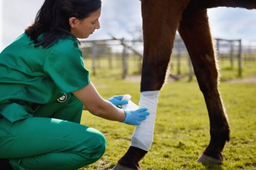
Following treatment, the surgeon closes the incisions in the skin using sutures. The surgical drapes are removed, and the limb is bandaged before the horse is aroused from general anesthesia.
Interpreting Results
Arthroscopy allows surgeons to visually assess a joint’s integrity, rather than relying on other diagnostic tools that may not reveal the true extent of damage.
Features of the joint they observe include: [2]
- Joint fluid
- Joint membrane
- Joint capsule and ligaments
- Cartilage and bone
Joint Fluid
Joint fluid is typically clear, yellow-tinged, and viscous (thick). It should not contain any debris or fragments. [2]
During arthroscopy, red-tinged joint fluid can indicate recent or reoccurring tissue disruption, resulting in bleeding into the joint space. Fragments or debris may indicate breakdown of the cartilage or bone. [2]
Joint Membrane
The synovium (joint membrane) produces joint fluid to ensure proper lubrication of the joint. It has characteristic villi (finger-like projections) on its inner surface. [2] Synovial villi react to damage within the joint by changing their color, density, and thickness. Surgeons can interpret these changes to make a diagnosis.
Common changes in the synovium include: [2]
- Hyperemia: Reddening of the villi indicating inflammation.
- Petechiation: Pinpoint red dots on the villi where small blood vessels have enlarged, indicating inflammation.
- Thickening: Swelling of the villi due to edema (fluid accumulation), suggestive of inflammation.
- Density changes: Coalescing or clumping of the villi can be a response to inflammation, as the body attempts to produce more joint fluid to maintain lubrication. Damaged villi may also develop scar tissue, forming dense mats.
Joint Capsule & Ligaments
The joint capsule is a layer of connective tissue that contains the joint fluid. Areas of high strain thicken to form a ligament, a more robust cord of connective tissue within the joint capsule. [2]
The joint capsule or its associated ligaments may tear or rupture, which can be identified on arthroscopy in some cases. [2]
Cartilage & Bone
The bones making up each joint are lined with a layer of cartilage, which helps provide a smooth, low-friction surface for joint movement. Arthroscopy allows the surgeon to evaluate the cartilage and edges of the bones to identify any changes.
Common findings include: [1][2]
- Osteophytes: Areas of bone proliferation at the edge of a joint, indicating arthritis.
- Enthesophytes: Bone proliferation where a ligament or tendon inserts onto the joint, indicating inflammation, previous ligament damage, and/or arthritis.
- Fibrillation: Irregularity in the cartilage surface producing waves, small craters, or linear erosions. Often indicates that there is a loose fragment within the joint space that is interfering with joint movement.
- Eburnation: Areas where the cartilage is completely worn away, revealing underlying bone. This typically results from chronic arthritis.
- Fragments: Can result from osteoarthritis or osteochondritis dissecans. These pieces break off from the bone or cartilage and float within the joint fluid, interfering with joint movement and increasing friction.
Preparing Your Horse for Arthroscopy
There are no specific steps needed to prepare your horse for arthroscopy, as most of the preparation is handled by the veterinary team once your horse arrives at the surgical center. Your veterinary surgeon can walk you through the preparation steps they undertake for your horse’s specific procedure.
After the procedure, your horse will have a postoperative care plan to ensure appropriate healing. The extent of aftercare depends on the joint involved, invasiveness of the procedure, and other factors. [1]
Common components of postoperative care for arthroscopy include: [1]
- Bandaging
- Anti-inflammatory medications
- Antibiotics
- Suture removal
Depending on surgeon preference, available facilities, and other factors, the initial recovery period may occur at the clinic or at home. Some surgeons may refer you to surgical recovery boarding facilities or rehabilitation facilities for ongoing management.
If your horse has undergone treatment, your surgeon may recommend a rehabilitation program to bring your horse back into work once their surgery site heals. They may also recommend intra-articular (into the joint) treatments for several weeks to help boost cartilage healing and reduce inflammation. [1]
A rehabilitation plan after arthroscopy may involve: [4]
- Stall rest with hand grazing only for up to 2 weeks
- Gradually introducing and increasing hand-walking exercise over a period of 6 weeks
- Turnout in a small paddock and a gradual return to under saddle work
During the recovery period, you should monitor for any swelling, discharge from the surgical site, fever, or lameness. Contact your horse’s surgical team if there are any signs of pain or other complications. [4]

Complications
Although arthroscopy is a safe and effective procedure, it comes with a risk of intraoperative (during the surgery) and postoperative (during recovery) complications. Significant complications are rare for this procedure.
Intraoperative Complications
The most common intraoperative complications in arthroscopy are: [5]
- Iatrogenic damage
- Foreign material in the joint capsule
Iatrogenic Damage
Iatrogenic damage refers to tissue injury caused by the surgeon during the surgery. It most commonly occurs due to aggressive technique, movement of the horse during surgery due to inadequate sedation or anesthesia, or poor visualization of the tissue during the procedure. [6]
If iatrogenic damage is identified, the surgeon treats the injury and reports the damage to the client. The prognosis depends on the extent of the damage, post-operative care, and other factors. To prevent iatrogenic injury, surgeons are trained in gentle techniques and optimal tool handling. They also ensure appropriate sedation/anesthesia during the procedure.
Foreign Material
Foreign material within the joint capsule can occur due to breakage of instruments or from pieces of drape, hair, skin, or adhesive entering the surgical incision. [5] In most cases, these fragments can be flushed out of the joint during the surgery.
Surgical instruments can break accidentally from interference between tools within the joint space or from movement of the horse during the procedure. Surgeons are trained in tool handling techniques to minimize the risk of interference. If an instrument does break, surgeons use endoscopy, radiographs, or ultrasound to locate the fragment for removal. [6]
Postoperative Complications
Postoperative complications associated with arthroscopy include: [5]
- Neuropathy or myopathy
- Surgical site infection
- Synovitis
Neuropathy or Myopathy
Following general anesthesia, horses may be unable to use their limb or limbs properly due to damage to the muscles or nerves. [6] Typically these conditions result from inadequate padding on the surgical table causing pinching or compression of the tissue. [6]
Prognosis depends on the severity of the injury. Most horses regain normal function within 10-14 days after the procedure. If there are still deficits present, such as difficulty moving a limb, after 14 days, the prognosis for full return to function is poor. [6]
Surgical Site Infection
Surgery creates the opportunity for bacteria to enter the body, potentially resulting in infection. To reduce the risk of infection, surgeons use aseptic technique to minimize the risk of bacteria entering the wound. They may also place the horse on antibiotics after the surgery to prevent infection from developing. [6]
The prognosis for surgical site infections depends on the location of the infection. Infections in the skin typically resolve rapidly with appropriate treatment.
Septic arthritis, bacterial infection of the joint, is a more severe complication that requires intensive treatment. [6]This condition is uncommon after arthroscopy, occurring in around 1% of surgeries. [7] Treatment for this condition may involve antibiotics, flushing of the joint, regional limb perfusion, and more. [5]
The prognosis for this condition can be poor, as bacterial infection causes significant damage to the joint tissues.
Synovitis
Some horses undergoing arthroscopy develop joint swelling after surgery. This swelling may last for more than 6 weeks. [6]
Synovitis usually indicates there is still disease present within the joint that was not completely treated by the procedure. [6] It can also develop if the horse resumes activity too quickly after surgery, resulting in low-grade inflammation within the joint. However, some horses develop idiopathic (unknown cause) synovitis that may persist for weeks to months. [6]
Treatment typically involves repeating the arthroscopy to remove any areas of diseased tissue that were missed or anti-inflammatory medication to reduce local inflammation. Idiopathic synovitis may resolve over time as the horse’s joint capsule reduces its increased production of joint fluid in response to the surgery. [6]
Prevention strategies include anti-inflammatories after the surgery and a gradual return to exercise.
Frequently Asked Questions
Here are some frequently asked questions about arthroscopy in horses:
Arthroscopy in horses is a minimally invasive surgical procedure that allows a veterinarian to look directly inside a joint using a small camera. The camera sends real-time images to a monitor so the surgeon can assess cartilage, bone, ligaments, and other joint structures. Small instruments can also be inserted through separate tiny incisions to treat problems during the same procedure. This makes arthroscopy useful for both diagnosis and treatment.
Arthroscopy is performed in horses to diagnose and treat joint problems that cause lameness, pain, or reduced performance. It is commonly used for conditions such as osteochondritis dissecans, synovitis, cartilage or bone fragments, ligament injuries, and some forms of osteoarthritis. In many cases, other tests strongly suggest the problem, but arthroscopy allows the surgeon to confirm exactly what is happening inside the joint. It also gives the surgeon a chance to remove damaged tissue or loose fragments right away.
Arthroscopy offers several benefits compared to traditional surgery because it uses small incisions rather than a larger open approach. This usually means less soft tissue trauma, less postoperative pain, a lower infection risk, faster healing, and less scarring. It also allows direct visualization of the inside of the joint, which can improve diagnostic accuracy. For many horses, these advantages support a smoother recovery and better long-term joint outcomes.
Arthroscopy is usually performed in horses under general anesthesia in a surgical setting. After the skin is clipped or shaved and carefully disinfected, the surgeon makes a small incision near the joint and inserts the arthroscope. Fluid or gas is used to expand the joint space and improve visibility, and a second incision may be made for surgical instruments. Once the joint has been examined and treated as needed, the incisions are closed with sutures and the limb is bandaged.
Arthroscopy can diagnose several kinds of joint problems in horses by allowing the surgeon to inspect the joint directly. It can help identify inflammation of the joint lining, cartilage damage, loose bone or cartilage fragments, ligament injuries, and changes associated with osteoarthritis. It can also reveal abnormalities in the joint fluid, joint capsule, and synovial tissues that may not be obvious on imaging alone. This direct view is especially helpful when other diagnostic tests are inconclusive.
Several treatments can be done during arthroscopy, depending on what the surgeon finds inside the joint. Common procedures include removing bone or cartilage fragments, debriding damaged tissue, flushing the joint to remove inflammatory or infectious material, and reconstructing parts of the joint surface. These treatments are designed to reduce friction, improve joint function, and support healing. Because the surgeon can both diagnose and treat the problem during the same procedure, arthroscopy is often very efficient.
Preparing your horse for arthroscopy usually does not require many special steps at home because most of the preparation is handled by the veterinary team at the surgical facility. Your surgeon will explain the plan for anesthesia, skin preparation, and the procedure itself based on the joint being treated. Owners are usually most involved in planning transportation, aftercare, and the recovery environment ahead of time. It is helpful to ask in advance what kind of stall rest, bandaging, and follow-up care will be needed after surgery.
Recovery after arthroscopy in horses usually involves bandaging, anti-inflammatory medication, and a carefully controlled return to movement. Many horses start with stall rest and limited hand grazing, then gradually add hand-walking over several weeks before progressing to small paddock turnout and eventually riding. The exact schedule depends on the joint involved, the amount of treatment performed, and how well the horse is healing. Close monitoring during recovery is important to catch swelling, discharge, fever, pain, or renewed lameness early.
The risks of arthroscopy for horses include both surgical and postoperative complications, although significant problems are uncommon. Potential issues include accidental tissue injury during surgery, foreign material entering the joint, nerve or muscle damage related to anesthesia, infection, and persistent joint inflammation. Septic arthritis is one of the more serious complications, but it is uncommon after arthroscopy. Overall, the procedure is generally considered safe when performed by an experienced surgical team.
Owners should watch for swelling, discharge from the incision site, fever, worsening lameness, or clear signs of pain after arthroscopy. These changes can suggest infection, persistent inflammation, or another postoperative complication that needs veterinary attention. Some mild swelling can occur, but marked or prolonged changes should not be ignored. If anything seems abnormal during recovery, the horse's surgical team should be contacted promptly.
Many horses can return to work after arthroscopy, especially when the problem is identified early and treated effectively. The likelihood of a good outcome depends on the underlying joint disease, how much tissue damage was present, and how well the horse follows the rehabilitation plan. Some horses need only a gradual conditioning program after healing, while others may need additional joint treatments during recovery. Long-term success is usually best when the joint is protected carefully during the return-to-work period.
Arthroscopy should be considered for a lame horse when a veterinarian suspects joint disease that needs direct evaluation or treatment. It is especially useful when imaging and examination suggest a problem such as osteochondritis dissecans, loose fragments, synovitis, or cartilage injury, but more detail is needed. In some horses, arthroscopy provides the definitive diagnosis after other tests leave uncertainty. It is most appropriate when knowing exactly what is inside the joint will change treatment or prognosis.
Summary
Arthroscopy is a minimally invasive surgical technique that allows veterinarians to diagnose and treat joint problems in horses.
- A small camera (arthroscope) is inserted into the joint to give the surgeon a clear, real-time view of cartilage, bone, and soft tissues.
- The procedure is commonly used to remove bone chips, address OCD lesions, treat synovitis, and repair cartilage damage caused by injury or wear.
- Arthroscopy involves smaller incisions than traditional surgery, leading to less pain and faster healing.
- This approach allows for both diagnosis and treatment in a single procedure, improving outcomes.
References
- Ross. M. W. and Dyson. S. J. Diagnosis and Management of Lameness in the Horse. 2nd ed. Elsevier Saunders, St. Louis, Mo. 2011.
- MacIlwraith. C. W. et al. Diagnostic and Surgical Arthroscopy in the Horse. Fourth edition. Mosby, St. Louis. 2015.
- Abutarbush. S. and Carmalt. J. Equine Endoscopy and Arthroscopy for the Equine Practitioner. Teton NewMedia, New York. 2008.
- Gasiorowski. J. C. and Richardson. D. W. Diagnostic and Therapeutic Arthroscopy in the Standing Horse. Veterinary Clinics of North America: Equine Practice. 2014.
- Goodrich. L. R. and McIlwraith. C. W. Complications Associated with Equine Arthroscopy. Veterinary Clinics of North America: Equine Practice. 2008.
- Rubio-Martinez. L. M. and Hendrickson. D. A. Complications in Equine Surgery. Wiley Blackwell, Hoboken. 2021.
- Olds. A. M. et al., Evaluation of the Rate of Development of Septic Arthritis after Elective Arthroscopy in Horses: 7 Cases (1994–2003). Journal of the American Veterinary Medical Association. American Veterinary Medical Association. 2006.