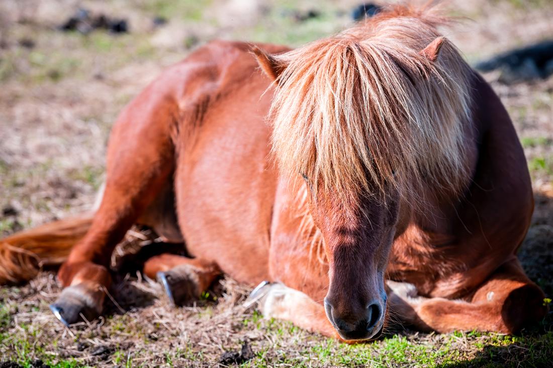
Acute laminitis refers to the first few days of a laminitis episode during which clinical signs are observed. Laminitis is a painful condition that causes damage to the hoof laminae, which anchor the coffin bone to the hoof wall.
During the acute phase, horses typically display signs of pain including a “rocked back” stance, a stiff gait, or a reluctance to move. The hooves may feel hot with a stronger digital pulse.
Left untreated, acute laminitis can cause life-threatening debilitation or lead to euthanasia. However, with prompt and aggressive treatment, most horses recover from the condition and return to soundness within two months. [1]
There are multiple and interrelated factors involved in the development of acute laminitis. Factors currently being investigated include inflammation, enzyme activation, insulin resistance, vascular endothelial dysfunction, and excessive weight-bearing on the hoof due to a severe lameness in the opposite limb.
Treatment for laminitis focuses on nutritional and medical management. Types of treatments include cryotherapy, anti-inflammatory therapy, pain management, and biomechanical interventions. The single most important part of treatment is to identify and remove the cause.
What is Acute Laminitis?
Laminitis affects the epidermal (insensitive) and dermal (sensitive) laminae of the equine hooves. It can occur in one or more hooves but is most common in the front hooves.
Laminitis can affect adult horses and ponies of any breed or age. However, horses with systemic illness or underlying endocrine diseases, including pituitary pars intermedia dysfunction (PPID) and equine metabolic syndrome (EMS), have an increased risk of this condition. [2]
The acute phase of laminitis involves the onset of clinical signs including pain, heat, and increased digital pulse. This phase can progress to the point that the coffin bone becomes displaced within the hoof capsule, known as founder.
Laminitis can become a chronic condition for some horses. Once a horse has had a bout of acute laminitis, they have an increased risk of future recurrence. [24]

Phases of Laminitis
There are five phases of laminitis recognized by veterinarians. These phases include the developmental phase, acute phase, subacute phase, chronic phase and refractory phase.
Developmental phase: The horse is exposed to one or more predisposing factors that trigger laminar separation in the hoof but with no outwardly visible signs of pain. This phase can last for anywhere from 8 to 60 hours depending on the triggering factor. [20]
Acute phase: The horse displays clinical signs of pain or lameness, along with a bounding digital pulse and heat in the hooves. This phase lasts between 24 to 72 hours and may conclude with the coffin bone rotating and sinking in the hoof, known as digital collapse. [20]
Subacute phase: If there is no evidence of coffin bone rotation or digital collapse after 72 hours of the acute phase, the horse is considered to progress to the subacute phase of laminitis. During this phase, the horse experiences less severe clinical signs and the hoof begins to recover. [20]
Chronic phase: When the coffin bone rotates and sinks (displacement of the distal phalanx), the horse progresses to the chronic phase of laminitis. This phase can last for a few months or it can last for the remainder of the horse’s life. Clinical symptoms may resolve during this period, or the horse may remain lame and continue to experience ongoing pain. [20]
Refractory phase: In some cases, the horse does not respond to conventional laminitis treatment within 7-10 days after the onset of the acute phase. These horses may have extensive damage to the laminae and severe pain. They may require surgical treatment and may never return to soundness. [20][21]

Types of Laminitis
Acute laminitis can develop due to a number of triggering factors, including metabolic dysfunction, as a secondary result of illness, following the use of certain medications, trauma to the hoof, and in support limbs when severe lameness is present.
The strategies used to resolve laminitis will vary depending on the initial cause of the condition. Some common causes of laminitis include:
Endocrinopathic laminitis: Occurs in association with endocrine conditions, including EMS and PPID. This type of laminitis may be triggered by a high intake of lush pasture (pasture-associated laminitis). It is mediated by high insulin levels. [3][4]
Illnesses that cause a systemic inflammatory response: Several illness are believed to associated with elevated toxins and or activation of enzymes that cause destruction of the basement membrane of the laminae, including:
- Colic
- Colitis
- Grain overload
- Strangles
- Lyme disease
- Potomac Horse Fever
- Retained placenta
- Pneumonia
Laminitis caused by bedding on black walnut shavings appears to share the same mechanism.
Supporting limb laminitis (SLL): The least common type of laminitis, SLL occurs in horses suffering a non-weight-bearing lameness. Laminitis develops in a supporting limb that is bearing more weight than normal.
Impaired blood supply in the foot: Poor blood flow to the feet can often cause laminitis. For example, horses that have one or more legs trapped in wire fence, etc. to the point that they have no or extremely compromised circulation often develop laminitis. Impaired circulation is known component of endocrinopathic laminitis.
Ingestion of some anti-nutritional factors: Some components of a horse’s diet might induce hoof health issues. For example, endophyte-infested tall fescue can cause laminitis. [26][27] In addition, low oxygenation of the blood can occur in Red Maple poisoning or from consumption of forages with high nitrate levels and cause laminitis-like hoof pain.
Clinical Signs of Acute Laminitis
Depending on the severity of a laminitis attack, horses can display a range of signs. Common signs of laminitis include:
- Lifting the hooves alternately and incessantly to shift the body weight from leg to leg
- Increased digital pulse in affected hooves
- Heat at the coronet band
- Mild to severe lameness
- Resistance to move
- Shuffling walk with head held either abnormally high or low and rigidly
- Short-strided gait or other gait abnormalities
- Standing with the front legs positioned in front of the body (rocked back stance)
- Muscular tension through the shoulders, back and rump
- Spending more time down
Diagnosis of Acute Laminitis
Veterinarian assessment is required to accurately diagnose laminitis. This assessment will involve reviewing past medical history, completing a medical evaluation, and potentially taking x-rays to determine if any displacement of the coffin bone (distal phalanx) has occurred.
Your veterinarian will also conduct a lameness exam to determine the severity of the case. Lameness is typically scored on the following 5-point scale: [18]
AAEP Lameness Scale
Grade 0: Lameness is not perceptible under any circumstances.
Grade 1: Lameness is difficult to observe and is not consistently apparent, regardless of circumstances.
Grade 2: Lameness is difficult to observe at a walk or when trotting in a straight line but is consistently apparent under certain circumstances.
Grade 3: Lameness is consistently observable at a trot under all circumstances.
Grade 4: Lameness is obvious at a walk.
Grade 5: Lameness produces minimal weight bearing in motion and/or at rest or a complete inability to move.
Treatments for Acute Laminitis
Acute laminitis is a medical emergency and should be treated based on the advice of a veterinarian.
The goals of treatment are to eliminate or minimize factors that triggered the condition, address pain, reduce or prevent damage to the laminae, and avert displacement of the coffin bone within the hoof capsule.
Treatment for acute laminitis typically includes a combination of the following strategies:
1) Dietary Management
Hyperinsulinemia in EMS or PPID is the cause of 90% of laminitis cases. If the horse develops laminitis with no other obvious cause, it should be presumed they have high insulin until blood work can rule it out.
Horses experiencing a bout of acute laminitis should be fed a diet that is low in hydrolyzable carbohydrate (HC) which is ethanol-soluble carbohydrates (ESC) and starch. Forage should be the predominant component of the diet and hay with less than 10% HC should be selected. This will limit the insulin response to feeding.
Hay analysis is strongly advised to determine the HC level and minerals so that a balanced diet can be developed.
Until the safety of the hay is determined, hay should be soaked for horses with acute laminitis. Soaking hay can significantly reduce the sugar content of the forage. It is recommended to soak hay for at least 30 minutes in warm water or 60 minutes in cold water. [25]
Avoid feeding concentrated feeds and commercial grain products, unless they are specifically designed to be low-carbohydrate rations with less than 10% HC. Horses should have no access to grass pasture during the acute phase of laminitis.
While you are waiting for your hay analysis results, you can implement the following supplementation protocol recommended by the ECIR group: [34]
- Use a small amount of thoroughly rinsed beet pulp to carry supplements and medications
- Provide 60 mL of w-3 oil
- Supplement 1,000 IU of vitamin E
- Supplement with 10 grams of magnesium oxide to provide roughly 5 grams of magnesium
2) Disease Management
Hormonal imbalances associated with Equine Metabolic Syndrome (EMS) and Pituitary Pars Intermedia Dysfunction (PPID) are directly associated with an increased risk of laminitis.
Horses with PPID may have higher levels of hormones including ACTH, cortisol, and other pituitary hormones, which can promote insulin resistance and laminitis. [5] PPID and concurrent insulin resistance require treatment with medication (such as Pergolide) to reduce the risk of laminitis.
Research conducted at a Finnish veterinary hospital on horses with laminitis found that 89% had evidence of underlying endocrine disorders. [2] One-third had a diagnosis of PPID and the remainder had high insulin levels without PPID indicative of metabolic syndrome. [2]
Insulin resistance is often present in overweight or obese horses. [6][7][8] Managing acute laminitis associated with equine metabolic syndrome requires controlling weight by restricting carbohydrate intake and following an appropriate exercise program.
Supplemental thyroid hormone is often prescribed to hasten weight loss. Horses with acute metabolic laminitis may be prescribed medication of metformin or one of the SGLT2 inhibitor drugs, ertugliflozin or canagliflozin, to get insulin under rapid control.
3) Cryotherapy
Cooling the hooves via cryotherapy (cold therapy) may help to reduce the damaging effects of laminitis on the laminae. It is recommended to cool the hoof wall surface to temperatures between 5°C to 7°C continuously for at least 48 hours.
Cryotherapy may prevent lameness, reduce the release of damaging enzymes within the laminae, and reduce inflammation within the laminae of horses with laminitis related to a systemic inflammatory response such as grain overload, experimental fructan overload or black walnut poisoning. [9][10][11][12][13]
A research study investigating cryotherapy for laminitis determined that horses with colitis were ten times less likely to develop laminitis when their hooves were cooled continuously for at least 48 hours. [14]
When to use ice
It should be noted that prevention is only possible when icing is started during the developmental phase, before the horse is lame. If started at the first sign of lameness, icing improved the outcome and reduced damage in horses with inflammatory laminitis in one study. [13]
Icing during the developmental stage also helps with metabolic laminitis but it is very difficult to predict when laminitis will occur unless there has been some dietary indiscretion like the horse breaking out and eating grass.
Icing after lameness has occurred has not been studied and may not be advisable because there is a strong component of vascular constriction in endocrinopathic laminitis and icing would make that worse.
How to apply ice
Cryotherapy is typically well tolerated by horses but is labour-intensive. To maintain the surface of the hoof wall at temperatures below 10°C, ice must be replaced approximately every one to two hours depending on the temperature of the surrounding environment.
For optimal results, cryotherapy should be applied to the hoof, pastern, fetlock, and a portion of the cannon bone. Using vinyl boots or plastic trash cans filled with ice and water or a circulating refrigerated bath are effective methods for administering cryotherapy.
Five-litre intravenous fluid bags can be filled with an ice slurry to use for cryotherapy and are large enough to cover the hoof and lower pastern. Gel packs do not provide sufficient cooling for the hooves.
4) Medications
Anti-inflammatory Drugs
Inflammation present during the early stages of acute laminitis caused by a systemic inflammatory response can contribute to the destruction of the laminae.
Endocrinopathic laminitis does not have the invasion of white cells typical of inflammation, or activation of destructive enzymes. [29] There may be some inflammatory reaction as a clean-up response to dead or damaged tissue in severe cases but anti-inflammatory drugs typically are not very effective in this type of laminitis because they do not address the root cause.
Anti-inflammatory medications are beneficial for horses with acute laminitis caused by a systemic inflammatory response by reducing laminar damage.
Phenylbutazone is a non-steroidal anti-inflammatory drug and is considered one of the most effective pain-relieving medications used in horses. Other anti-inflammatory medications used include Firocoxid and Flunixin Meglumine – a drug that is administered intravenously.
Dimethyl sulfoxide (DMSO) is an anti-inflammatory drug that is applied topically to the coronet bands of horses affected by laminitis. In some cases, the drug is administered intravenously.
MMP Inhibitors
Some horses develop laminitis in response to a compromised intestinal lining caused by hindgut acidosis, ischemia (lack of blood flow) or bacterial infection. When the intestinal barrier is not functioning properly, more toxins will be absorbed from the gut and enter the bloodstream.
These circulating toxins activate enzymes in the laminae known as matrix metalloproteinases (MMPs), which damage the hoof laminae and contribute to laminitis. [15][16]
Medications that inhibit the destruction of laminar connections due to MMPs are used in some cases of acute laminitis. Batimastat was a MMP inhibitor developed for laminitis but is no longer available due to poor response.
Pentoxyfylline and doxycycline are indirect inhibitors of MMP because of their anti-inflammatory properties. Like icing, MMP inhibition likely needs to be done during the developmental stage to be really effective.
Note that endocrinopathic laminitis does not involve MMP activation.
Analgesics
Phenylbutazone (‘bute’) is a nonsteroidal anti-inflammatory drug and the most common drug used for treating laminitic pain in horses. All drugs in this class should be used at the lowest dose possible and for no longer than 7 days to avoid gastric, colonic and kidney side effects. Bute and other NSAIDs do not work as well in endocrinopathic laminitis.
In some cases of acute laminitis, horses need additional analgesic (pain-relieving) medications administered.
Lidocaine, Ketamine, and Butorphanol are pain management medications that are administered intravenously in a hospital setting. Gabapentin can be administered orally or intravenously, and Morphine can be administered intramuscularly or intravenously. These medications should only be used under veterinary guidance.
Tramadol is a good choice for outside the hospital setting. [30]
5) Limiting Movement
Minimizing additional stress on the hoof laminae requires restricting movement while horses are recovering from laminitis. Confining the horse to a well-bedded stall or small area with soft footing is ideal to prevent movement that could promote further injury to the laminae.
However, stall confinement limits circulation which is often already impaired. Allow your horse turnout in a small area or area once the horse is comfortable, willing to walk and off pain medications.
6) Hoof Care to Improve Biomechanics
Getting proper hoof care is often the most difficult thing for owners to achieve but it is critical to comfort and recovery. There are almost as many opinions on how to deal with laminitic hooves as there are horses with hooves.
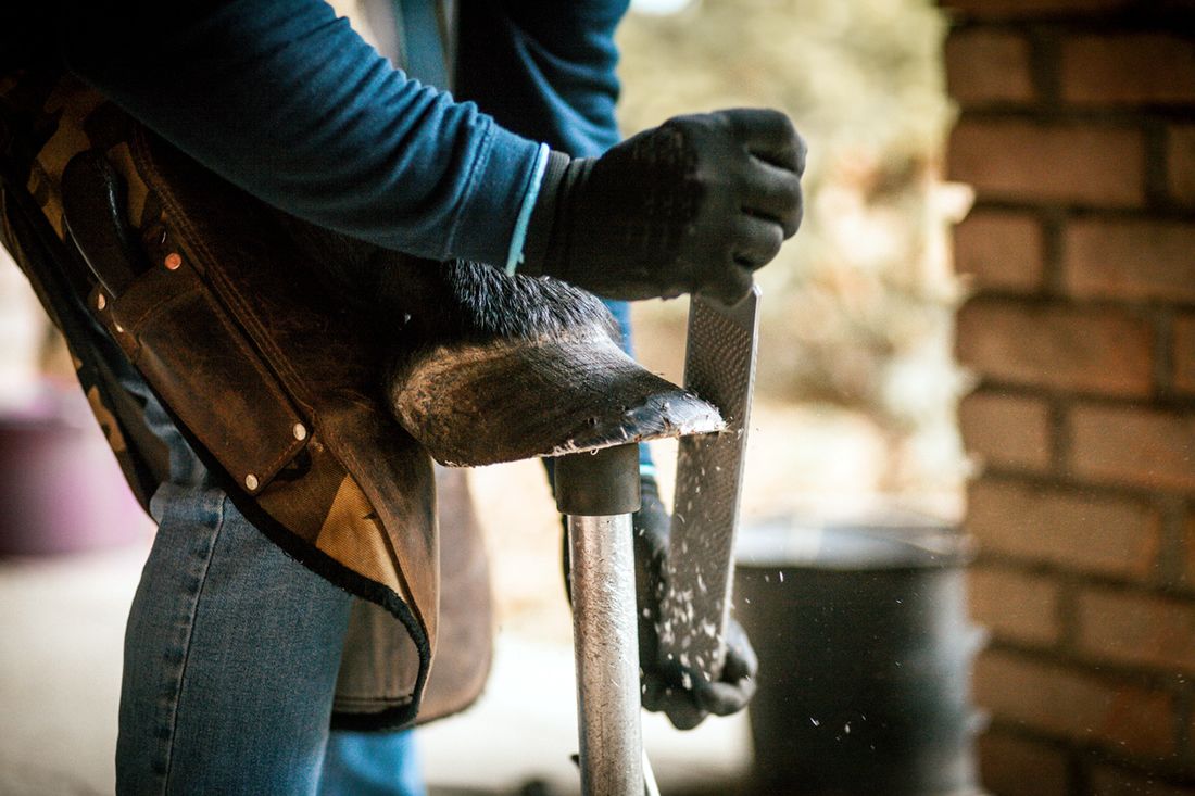
As soon as you recognize laminitis in your horse, contact your vet to get radiographs (x-rays) done and connect with your farrier to achieve a physiologically correct trim.
An x-ray will reveal the position of the coffin bone and whether any rotation has occurred. The following features are typical of a normal hoof:
- Same distance between:
- Edge of the coffin bone to the hoof wall
- Tip of the coffin bone to the tip of the toe
- Bottom of the coffin bone to the ground with a 5o palmar angle between the ground and coffin bone
- On the sole surface, the back of the heels is level with the widest part of the frog
In this ideal foot, at least 2/3 of the ground surface is in the back of the foot. The sole, frog, and internal digital cushion are doing most of the shock absorption.
With laminitis, the connection between the hoof wall and coffin bone is weak. The last thing you want to do is put all the weight on the walls. Ideally, the walls are beveled back to the level of the laminae.
Styrofoam pads are probably the most commonly used appliance for acute laminitis. [17] They are usually taped on, allowed to crush down then the front half cut off. The back is applied to a new pad underneath before taping them on, to provide good support at the frog and heel.
Optimal trim interval can be as short as 2 weeks. Leaving the horse barefoot in pads and shoes is the best approach because frequent trims may be needed to achieve the desired foot. Laminitic feet tend to grow faster, sometimes much faster at the heels.
A variety of shoes and other devices are used for chronic laminitis but even they have the basic realigning trim as a mandatory starting point. The only shoe that has been proven to limit the movement of the coffin bone in laminitic horses is the heart bar shoe. [31] This requires skill and hot forging to get the correct fit for the individual horse.
Prevention of Acute Laminitis
If your horse is showing early warning signs of laminitis, contact a veterinarian to confirm the diagnosis and develop a treatment plan.
You can reduce your horse’s risk of acute laminitis and prevent future flare-ups by implementing the following diet and management practices:
1) Feed a Diet Low in Hydrolyzable Carbohydrates
Avoid feeding a diet with high levels of HC (starch + simple sugars) to horses at risk of metabolic syndrome. This includes limiting or eliminating concentrate feeds and choosing a low-HC forage. Diets high in starch and sugar increase blood sugar and insulin levels.
Research shows that horses many young horses with EMS resolve laminitis within 2 weeks when placed on a safe, hay only diet and their insulin level rapidly normalizes. However, there is a subpopulation of EMS horses with more severe disease, higher insulin and glucose, which do not respond as well to diet alone. [19]
A low sugar, low starch, forage-based diet will support your horse’s overall well-being and metabolic health.
2) Provide Balanced Nutrition
Ensure your feeding program provides all the essential nutrients required to support hoof growth. Key nutrients for healthy hooves include:
Amino acids: Hoof tissue is composed of the protein keratin. Amino acids – such as lysine, methionione and threonine – are the building blocks pf protein necessary for hoof growth.
Biotin: A B-vitamin that is required for keratin production. Feed a minimum of 20 mg per day of biotin to support hoof health.
Minerals Microminerals including copper and zinc help to form the structural tissue that makes up the hoof. Iron overload has been identified in a variety of animals, including horses. [32] In humans, high body iron is also a risk factor for metabolic syndrome and insulin resistance increases iron absorption. [33]
Mad Barn’s AminoTrace+ vitamin and mineral supplement is specifically designed for horses at risk of laminitis. AminoTrace+ provides balanced levels of key nutrients, with no added iron, for hoof health and metabolic function.
3) Monitor Your Horse’s Weight
Ensure your horse maintains a healthy weight by regularly monitoring body condition and adjusting their diet and exercise plan accordingly.
Easy weight gain starting when the horse reaches physical maturity or if exercise stops is a sign of metabolic syndrome.
Horses that have recently gained weight and are overweight or obese have a four times greater risk of developing laminitis. [22] Horses that have excess fat deposits in regions such as the neck are also at an increased risk of laminitis as this is characteristic of metabolic syndrome. [23]
4) Provide Regular Hoof Care
Work with a farrier to have trimming/shoeing completed at regular intervals to maintain healthy hooves and facilitate proper movement. Some horses need protection from hoof boots and padding materials to help them move without discomfort.
5) Treat Metabolic Disease
Horses with insulin resistance and PPID should be treated promptly. These conditions contribute to hormonal imbalances that can impair normal cellular morphology and circulation in the hooves.
Summary
Keep the following guidelines in mind when feeding a horse with acute laminitis:
- Avoid all foods high in starch and sugar including grains, carrots, apples, untested hay/silage, molasses, and grass
- Feed low sugar and starch hay; soak the hay to reduce the sugar content
- Ensure a minimum of 1.5% bodyweight of forage is fed unless recommended otherwise by your veterinarian
- Replace up to 50% of the diet with straw; introduce this change gradually. Note: While straw is somewhat lower in calories than low HC hay, it is much lower in protein and minerals and may necessitate additional supplementation. Straw should also be checked carefully for residual grain and any molding.
- You may feed a very low sugar and starch unmolassed chaff, or well rinsed then soaked beet pulp as a carrier to ensure vitamins and minerals, supplements, and medications are eaten
- Feed an advanced probiotic supplement such as Optimum Digestive Health to support gut function
- Ensure salt and fresh water are available to your horse
- Ensure any metabolic conditions are well controlled
Mad Barn nutritionists can help you design a balanced feeding plan to support your horse’s recovery from acute laminitis. Submit your horse’s information online for a free consultation.
References
- Mitchell CF, et al. The management of equine acute laminitis. Vet Med (Auckl). 2014. View Summary
- Karikoski, N.P. et al. The prevalence of endocrinopathic laminitis among horses presented for laminitis at a first-opinion/referral equine hospital. Dom Anim Endocrin. 2011.View Summary
- Geor RJ. Pasture-associated laminitis. Vet Clin North Am Equine Pract. 2009. View Summary
- Geor RJ. Current concepts on the pathophysiology of pasture-associated laminitis. Vet Clin North Am Equine Pract. 2010. View Summary
- Donaldson, M.T. et al. Evaluation of suspected pituitary pars intermedia dysfunction in horses with laminitis. J Am Vet Med Assoc. 2004. View Summary
- Geor, R.J. Metabolic Predispositions to Laminitis in Horses and Ponies: Obesity, Insulin Resistance and Metabolic Syndromes. J Equine Vet Sci. 2008.
- de Laat, M.A. et al. Equine laminitis: induced by 48 h hyperinsulinaemia in Standardbred horses. Equine Vet J. 2010. View Summary
- Asplin, K.E. et al. Induction of laminitis by prolonged hyperinsulinaemia in clinically normal ponies. Vet J. 2007. View Summary
- Pollitt, CC. et al. Prolonged, continuous distal limb cryotherapy in the horse. Equine Vet J. 2004. View Summary
- van Eps, AW. et al. Equine laminitis: cryotherapy reduces the severity of the acute lesion. Equine Vet J. 2004.View Summary
- Van Eps AW. et al. Equine laminitis model: cryotherapy reduces the severity of lesions evaluated seven days after induction with oligofructose. Equine Vet J. 2009.View Summary
- van Eps AW. et al. Digital hypothermia inhibits early lamellar inflammatory signalling in the oligofructose laminitis model. Equine Vet J. 2012. View Summary
- van Eps AW. Et al. Continuous digital hypothermia initiated after the onset of lameness prevents lamellar failure in the oligofructose laminitis model. Equine Vet J. 2014. View Summary
- Kullmann, A. et al. Prophylactic digital cryotherapy is associated with decreased incidence of laminitis in horses diagnosed with colitis. Equine Veterinary Journal. 2014. View Summary
- Tadros, E.M. et al. Effects of a “two-hit” model of organ damage on the systemic inflammatory response and development of laminitis in horses. Vet Immunol Immunopath. 2012.View Summary
- Visser, M.B. and Pollitt, C.C. Lamellar leukocyte infiltration and involvement of IL-6 during oligofructose-induced equine laminitis development. Vet Immunol Immunopath. 2011.
- Goetz TE. The treatment of laminitis in horses. Vet Clin North Am Equine Pract. 1989. View Summary
- LAMENESS EXAMS: Evaluating the Lame Horse. AAEP.
- Sillence, M et al. Demographic, morphologic, hormonal and metabolic factors associated with the rate of improvement from equine hyperinsulinaemia-associated laminitis. Vet Clin North Am Equine Pract. 2010. View Summary
- Hembroff, D. Laminitis. Vetfolio.
- Hunt, RJ et al. Mid-metacarpal deep digital flexor tenotomy in the management of refractory laminitis in horses. Vet Surg. 1991. View Summary
- Wylie CE et al. Risk factors for equine laminitis: a case-control study conducted in veterinary-registered horses and ponies in Great Britain between 2009 and 2011. Vet J. 2013 View Summary
- Fitzgerald, DM. et al. The cresty neck score is an independent predictor of insulin dysregulation in ponies. PLoS One. 2019. View Summary
- de Laat, MA. et al. Incidence and risk factors for recurrence of endocrinopathic laminitis in horses. J Vet Intern Med. 2019. View Summary
- Bochnia, M. et al. Effect of Hay Soaking Duration on Metabolizable Energy, Total and Prececal Digestible Crude Protein and Amino Acids, Non-Starch Carbohydrates, Macronutrients and Trace Elements. J Equine Vet Sci. 2021.
- Rohrbach, B.W. et al. Aggregate risk study of exposure to endophyte-infected (Acremonium coenophialum) tall fescue as a risk factor for laminitis in horses. Am J Vet Res. 1995. View Summary
- Douthit, T.L. et al. The impact of endophyte-infected fescue consumption on digital circulation and lameness in the distal thoracic limb of the horse. J Anim Sci. 2012. View Summary
- Gauff, F. et al. Hyperinsulinaemia increases vascular resistance and endothelin-1 expression in the equine digit. Equine Vet J. 2013. View Summary
- Patterson-Kane, J.C. et al. Paradigm shifts in understanding equine laminitis. Vet J. 2018. View Summary
- Guedes, A. et al. Plasma concentrations, analgesic and physiological assessments in horses with chronic laminitis treated with two doses of oral tramadol. Equine Vet J. 2016. View Summary
- Aoun, R. et al. Shoe configuration effects on third phalanx and capsule motion of unaffected and laminitic equine hooves in-situ. PLoS One. 2023.
- Kellon, E.M. and Gustafson, K.M. Possible dysmetabolic hyperferritinemia in hyperinsulinemic horses. Open Vet J. 2019. View Summary
- Gonzalez-Dominguez, A. et al. Iron Metabolism in Obesity and Metabolic Syndrome
. Int J Molecular Sci. 2020. - Kellon, E.M. DDT +E – Diet. ECIR Group. Accessed November 6, 2023.


Leave A Comment