


Respiratory health plays a vital role in equine performance, comfort, and overall well-being. Horses are highly susceptible to a variety of lower airway conditions, many of which can present with subtle or nonspecific clinical signs such as intermittent coughing, reduced exercise tolerance, or nasal discharge. Accurate diagnosis of these conditions is essential to guide appropriate treatment and management.
One of the most informative and widely used diagnostic procedures for evaluating the lower respiratory tract in horses is bronchoalveolar lavage (BAL). This is a minimally invasive technique that allows veterinarians to sample cellular components from deep within the airways.
By introducing a small volume of sterile fluid into the lungs and recovering it for analysis, BAL provides direct insight into the inflammatory status of the lower airways. This information is particularly useful in diagnosing respiratory disorders such as equine asthma.
Read on to learn more about BAL, including indications, methodology, interpretation, and its clinical significance in equine practice. Whether used as a primary diagnostic tool or a method for monitoring disease progression and treatment response, BAL remains an essential component in the comprehensive assessment of equine lower airway disease.
Bronchoalveolar Lavage for Horses
Bronchoalveolar lavage (BAL) is a diagnostic technique used by veterinarians to examine the cell populations deep within the lung tissue. It involves passing a tube through the horse’s nostril into the airways, instilling a large volume of fluid, then removing the fluid and examining it under a microscope. [1]
Bronchoalveolar lavage specifically targets the deepest regions of the horse’s lungs. The veterinarian passes a narrow tube as far as possible into the lungs, lodging it within a small airway. Examining the collected fluid provides highly detailed information about the cell population within the lungs, allowing for a more accurate diagnosis.
Bronchoalveolar lavage helps veterinarians diagnose lower airway diseases, particularly: [2]
In some cases, veterinarians may perform a BAL when investigating poor performance in equine athletes. Repeated BAL procedures can also help monitor treatment efficacy in horses diagnosed with inflammatory respiratory diseases.
Clinical Use
Veterinarians use a bronchoalveolar lavage when they suspect lower airway disease, or conditions affecting the lungs and trachea (windpipe).
Signs of lower airway disease in horses include: [2]
- Coughing
- Nasal discharge
- Difficulty breathing, including rapid or very deep breathing
- A “heave line”- increased abdominal musculature from difficulty breathing
- Abnormal lung sounds when listening with a stethoscope
- Nostril flaring at rest
- Poor performance
- Exercise intolerance
Based on these findings, the veterinarian proceeds with diagnostic tests to determine the cause of lower airway disease.
Tracheal Aspiration vs. Bronchoalveolar Lavage
The two main tests used when investigating the airways are tracheal aspiration and bronchoalveolar lavage. Although both tests involve introducing fluid into the lungs, their key difference is the area of the lung sampled.
Tracheal Aspiration
During coughing, inflammatory cells, mucus, and bacteria are dislodged into the horse’s large airways. Tracheal aspiration (also referred to as Transtracheal Wash, or TTW) takes a sample from the “tracheal pool” which is towards the end of the trachea, allowing the veterinarian to assess if there is an infectious disease process occurring. However, the sample collected from the tracheal pool is not representative of what is collected from the deeper airways during a BAL.
When examining a sample of tracheal aspirate, the veterinarian is typically evaluating for bacterial infection by confirming the presence of bacterial colonies or other microbes. The presence of bacteria or other microorganisms on tracheal aspiration supports the diagnosis of infectious lower airway disease, like pneumonia. This test is not suitable for evaluating the number or proportion of bacteria cells present, as the size of the sampled area of lung tissue is unknown. [1]
Bronchoalveolar Lavage
In contrast, BAL collects a cellular sample from the lower airways. It is best for diagnosing non-infectious conditions that affect the lungs, like equine asthma or EIPH. [1]
By sampling a small portion of tissue of a known size, the proportion and number of cells present at that location can be quantified. This allows for more precise diagnosis of non-inflammatory conditions by determining which cells have had proportional increases or decreases. [1] Characterizing the types of cells present, as well as their relative numbers, allows for more precise diagnosis of non-infectious conditions.
What's your top priority with your horse's health?
Enter your email to receive your store credit
Equipment
There are two basic methods for performing a bronchoalveolar lavage: using a BAL catheter or an endoscope. [1] The method performed depends on the veterinarian’s preference, access to equipment, and whether the procedure is taking place in-clinic or on-farm.
To perform a BAL using a catheter, the veterinarian requires the following equipment: [1]
- Bronchoalveolar lavage catheter: A catheter around 2 – 3 meters in length
- Small empty syringe: To inflate the balloon that holds the catheter in place
- Local anesthetic: Numbs the airways and nasal passages
- Sterile saline in syringes: To introduce into the airway to collect cells
- Collection tubes: Stores the sample until it can be examined
When using an endoscope, the required equipment is similar. However, instead of using a bronchoalveolar lavage catheter, the veterinarian uses a 3-meter-long endoscope.
This procedure is typically performed under standing sedation, as most horses will not tolerate the procedure without significant chemical restraint. Veterinarians sedate the horse to prevent distress and excessive movement of the head. [1]
When possible, placing the horse in a set of stocks helps protect handlers and the veterinarian. In some cases, handlers may place a twitch on the horse’s muzzle to distract the horse from the procedure. [1]
Procedure
The bronchoalveolar lavage procedure begins with positioning the horse in a safe area, free from obstacles that may injure a panicking horse. Many veterinarians prefer to put the horse in stocks for this reason.
The horse is then sedated and left to stand quietly for a period of 5 – 10 minutes until the sedation is in full effect. [1]
1) Preparation
While the horse’s sedation comes into effect, the veterinarian prepares the catheter or endoscope. They check that the equipment is functioning properly and that it was properly sterilized since its last use. [1]
Next, the veterinarian administers 10 – 20 mL of local anesthetic into the horse’s nostril. [1] This numbs the tissue and helps reduce discomfort as the tube is passed through the nasal passage.
Once the local anesthetic has reached full effect, the handler typically places a twitch to help maintain control of the horse’s nose and provide a distraction. [1] The veterinarian then lubricates the tip of the catheter or endoscope and introduces it into the horse’s nostril.
The preferred head position for this procedure is with the head and neck extended as far as possible, to help the catheter or endoscope enter the horse’s trachea, rather than going down the esophagus. [3] A second handler may position the horse’s head, or a specialized halter may be used to hang the horse’s head from the stocks or ceiling. [3]
2) Sampling
As the veterinarian advances the catheter or endoscope, the horse may cough due to irritation in the respiratory tract. The veterinarian may administer additional local anesthetic to numb the respiratory tissues and help prevent coughing. [1]
When the catheter reaches the carina, the branching path of the trachea, the veterinarian inflates the balloon that holds the tube in place. [1] For endoscopes, a second handler typically maintains the position of the endoscope by holding it gently. [3]
 Illustration: Dr. Ana Mesa, PhD
After positioning the catheter or endoscope, the veterinarian introduces sterile saline into the horse’s lungs. [1] They then aspirate (draw back through suction) a syringe full of fluid and discard it. This first syringe contains the sterile saline that remained within the tube and did not reach the horse’s lungs. [1]
The veterinarian then slowly aspirates the remaining fluid out of the lungs into syringes. [1] These syringes contain the sample required for testing. At least 30 – 50 mL of sample should be acquired in a successful procedure. [1] A handler or assistant places this fluid into collection tubes.
Once the sample is acquired, the veterinarian carefully removes the endoscope or catheter. The handler removes the twitch and allows the horse to revive from sedation. Any sterile fluid not recovered during the procedure will be absorbed or expelled through coughing.
3) After Care
After the procedure, horses should not be exercised for 24 hours. [3] Very heavy exercise, such as galloping or jumping, should not be performed for 48 – 72 hours after the procedure. [3]
Horses should also be monitored closely for 48 hours or more for any signs of pneumonia, such as reduced appetite, increased respiratory effort, and fever. [2]

Interpreting Results
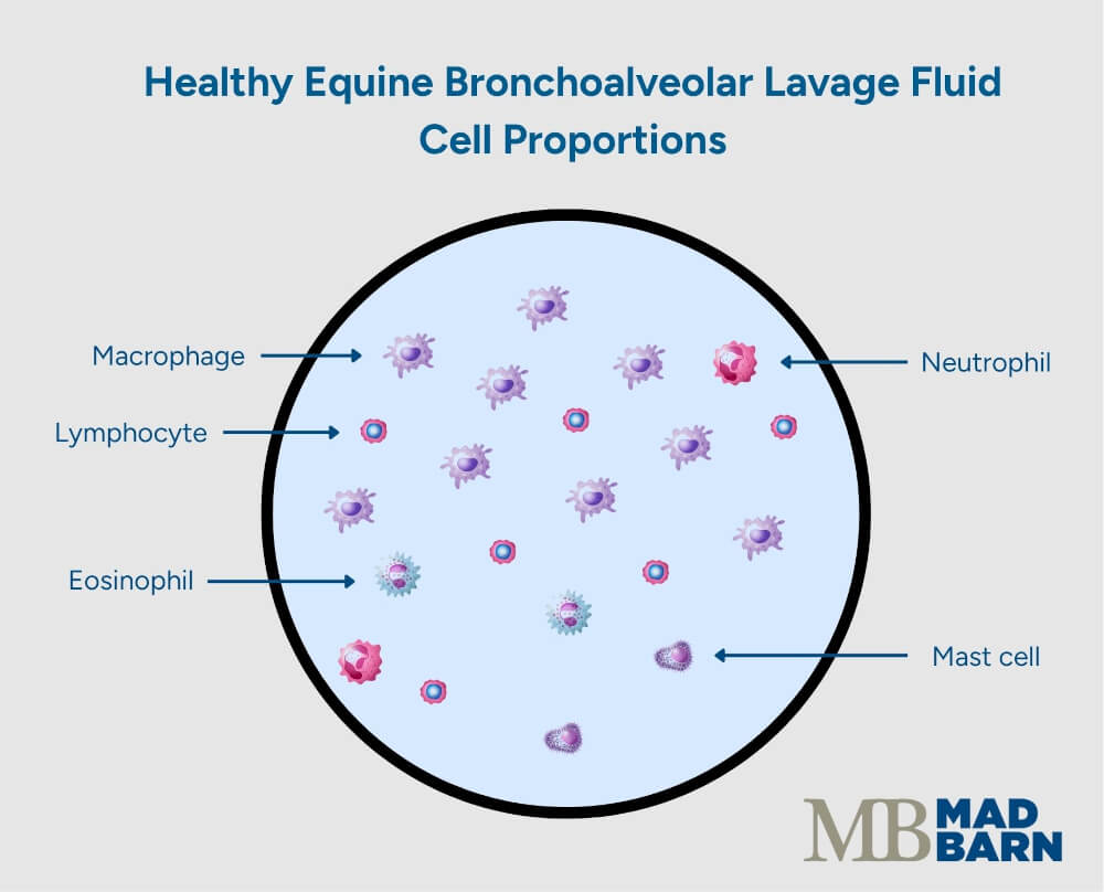
The collected sample is examined under a microscope, typically by diagnostic specialists such as veterinary pathologists. During the examination, the veterinarian looks for what types of cells are present and in what proportion.
Healthy equine BAL fluid contains the following cell proportions: [3]
- 50 – 70% macrophages: the body’s major clean-up cell
- 30 – 50% lymphocytes: the cells that produce antibodies
- Low numbers of neutrophils: cells that respond to bacterial infections
- Low numbers of mast cells and eosinophils: cells that respond to allergens
Changes in the proportions of these cells may indicate disease. There are three main conditions that a bronchoalveolar lavage can diagnose: equine asthma, pneumonia, and exercise-induced pulmonary hemorrhage.
Equine Asthma
Increased nondegenerate neutrophils on a BAL is an indicator of equine asthma. [3][4] Nondegenerate neutrophils are cells that have not been activated against a bacterial invader.
An increased population of these cells in the lungs indicates there is inflammation present, however the inflammation is sterile (not associated with bacteria). [4]
Some horses with equine asthma have increased mast cells and eosinophils, which may be associated with more severe disease and airway changes. [5]
Pneumonia
Horses with pneumonia show a high proportion of degenerate neutrophils in their BAL samples. These neutrophils have been activated against a bacterium, resulting in visible changes to the cell that the veterinarian can identify.
Common changes include karyolysis (degeneration of the cell’s nucleus) and pyknosis (fragmentation of the nucleus). [4]
The veterinarian can also use the acquired BAL sample for bacterial culture, a diagnostic test to identify the type of bacteria present in a sample. [6] In this procedure, the sample is placed on bacterial growth media and incubated to stimulate bacteria growth.
Based on the appearance of the bacterial colonies and what type of media they grow on, the veterinarian can determine the most likely bacterial species present. [6] This information aids diagnosis and guides the choice of antibiotic required to combat the infection.
Exercise-Induced Pulmonary Hemorrhage
Horses with exercise-induced pulmonary hemorrhage (EIPH) often have red blood cells within their BAL sample. They may also have very large macrophages that contain broken down red blood cells, indicating that the macrophages are cleaning up blood in the airways. [4]
Preparing for a BAL
Depending on your veterinarian’s preference, they may request that you bring your horse into their clinic to perform a BAL. In some cases, you may be referred to a specialist if your veterinarian does not have the appropriate equipment available to perform the procedure.
If your horse is nervous about trailer loading, you may need to practice loading in advance to ensure everything goes smoothly on appointment day.
There is no special preparation necessary for a BAL. Horses do not require a fasting period before the procedure.
If your horse is needle shy, your veterinarian may ask you to provide an oral sedative, such as Dormosedan® gel, one hour prior to the appointment. This medication takes at least 40 minutes to produce a sedative effect. It allows your veterinarian to provide additional sedating medications safely.
After the procedure, follow your veterinarian’s guidelines on resting your horse. Most veterinarians recommend resting your horse for at least 24 hours, and not performing intense exercise for up to 72 hours.
Complications
The bronchoalveolar lavage is a very safe procedure with minimal risk of complication. [7]
The most common complications associated with a BAL are fever, coughing, and reduced oxygen absorption leading to hypoxemia (low blood oxygen). [7] These conditions typically resolve within hours after the procedure. [7]
Rarely, more severe complications may develop. There are case reports of horses developing hemopneumothorax (blood and air in the thoracic cavity) or pneumonia after a BAL procedure. [2][7] In most cases, these conditions are treatable and have a good prognosis. [7]
Frequently Asked Questions
Here are some frequently asked questions about bronchoalveolar lavage in horses:
Bronchoalveolar lavage is a diagnostic procedure used to collect cellular samples from the lower airways of the lungs. It involves passing a tube into the horse’s airway and flushing a small amount of sterile fluid into the bronchi and alveoli, which is then retrieved for analysis.
BAL is commonly used to diagnose respiratory conditions such as equine asthma or exercise-induced pulmonary hemorrhage. It helps veterinarians evaluate the type and proportion of cells present deep within the lung tissue.
Yes, BAL is generally a safe and well-tolerated procedure when performed by an experienced veterinarian. Sedation minimizes stress and discomfort. Complications are rare, but can include mild coughing, nasal discharge, or temporary fever.
Receiving results from your horse's bronchoalveolar lavage depends on several factors, including shipping time to an appropriate diagnostic laboratory. Once the laboratory receives the sample, preliminary results from cytology are usually available within 1–3 days. Culture results, if requested, may take up to 7 days.
Summary
Bronchoalveolar lavage (BAL) is a diagnostic technique used to diagnose lower airway diseases in horses.
- Lower airway diseases often present with clinical signs such as coughing, nasal discharge, and poor performance
- The bronchoalveolar lavage procedure involves passing a tube into the horse's lungs, introducing sterile fluid and recovering it for examination
- The BAL is best for diagnosis of non-bacterial conditions, such as equine asthma and exercise-induced pulmonary hemorrhage
- BALs are a safe and effective method of diagnosing respiratory diseases
References
- Costa. L. R. R. and Paradis. M. R., Manual of Clinical Procedures in the Horse. Wiley Blackwell, Hoboken. 2018.
- Hewson. J. and Arroyo. L. G., Respiratory Disease. Veterinary Clinics of North America: Equine Practice. 2015.
- Dixon. C., Performing Bronchoalveolar Lavage in Horses in the Field. In Practice. 2022.
- Couetil. L. L. and Thompson. C. A., Airway Diagnostics. Veterinary Clinics of North America: Equine Practice. 2020. View Summary
- Karagianni. A. E. et al., Distinct Molecular Profiles Underpin Mild-To-Moderate Equine Asthma Cytological Profiles. Cells. 2024. View Summary
- Vitale. V. et al., Cytological Findings in Bronchoalveolar Lavage Fluid of Foals With Pneumonia Caused by Rhodococcus Equi and Other Bacteria. Journal of Equine Veterinary Science. 2019. View Summary
- Kopec. E. K. et al., Haemopneumothorax as a Complication of Bronchoalveolar Lavage. Equine Veterinary Education. 2024.


