


Electrocardiography (ECG) is one of the most important tools veterinarians use to evaluate heart health in horses. While stethoscopes remain invaluable for identifying murmurs and abnormal rhythms during a physical exam, an ECG provides a more precise and objective way to assess the heart’s electrical activity.
By recording and graphing the electrical impulses that control each heartbeat, an ECG allows veterinarians to detect rhythm disturbances, known as arrhythmias, that may not always be apparent on auscultation (i.e. using a stethoscope) alone.
Heart rhythm abnormalities are a common finding in horses and range from benign variations with little clinical impact to serious conditions that compromise performance or even endanger the horse’s life. An ECG helps determine whether an irregularity is harmless, requires monitoring, or needs treatment.
Although ECGs do not provide information about the physical structure of the heart, they are an important first step in diagnosing rhythm disturbances and guiding further testing, such as echocardiography. Keep reading to explore how ECGs are performed in horses, when they are most useful, and what information they provide for veterinarians and horse owners alike.
Equine Electrocardiogram (ECG)
Electrical signals control the horse’s heartbeat, ensuring consistent, coordinated beats for the entirety of the horse’s life. Improper electrical function can result in heart failure, poor performance, and even death.
An electrocardiogram (ECG; also known as EKG) is a diagnostic test that measures electrical signals across the heart, allowing veterinarians to diagnose arrhythmias. Arrhythmias are abnormal heartbeats resulting from irregular electrical signaling in the heart.
The ECG is a non-invasive procedure that is simple to perform and can provide insights into the horse’s heart function both at rest and during exercise.
Potential reasons why a horse might need an electrocardiogram include: [1]
- Arrhythmia detected on auscultation (listening with a stethoscope)
- Poor performance
- Exercise intolerance
- Pre-purchase examinations to confirm normal cardiac function
- Weakness or collapse
- Monitoring known structural abnormalities in their heart
- Monitoring treatment of arrhythmias
- Assessment prior to anesthesia
Depending on the condition the veterinarian suspects, they may request a resting or exercising ECG, or both. [1]
Typically, exercising ECGs are used for horses with a history of exercise intolerance or poor performance, as not all arrhythmias are detectable in the resting horse. [1] Performing an ECG during exercise highlights the abnormal electrical signaling and allows for diagnosis.
ECG Equipment
ECG devices are relatively simple pieces of equipment with a few key features: [1]
- Electrodes
- Recording device
- Tracing display
The electrodes are typically housed in clips or sticky pads that the veterinarian attaches to the horse’s skin. [1]
A liquid, such as water, saline, alcohol, or gel is applied to the pads or clips to improve contact between the horse’s skin and the metal components of the electrodes. This ensures adequate electrical conduction between the horse and the ECG machine. [1] Typically, clipping the ECG attachment sites is not necessary unless the horse has a full winter coat. [1]
The ECG recording device is typically a small computer that may also have a display for viewing the recorded tracing. Alternatively, the recording device may send its data to another computer or device to show the tracing. [1]
For exercise ECGs, the recording device is typically attached to the horse using a surcingle or girth attachment. The recording device can send a wireless signal to a viewing device, allowing the veterinarian to observe the ECG in real-time.
What's your top priority with your horse's health?
Enter your email to receive your store credit
Procedure
The process for applying an ECG is simple. The veterinarian sets up the ECG equipment, then identifies the locations where they want to place electrodes. The electrodes are placed in specific locations to detect electrical signals produced by the heart. The veterinarian applies gel, alcohol, saline, or water to the location, then attaches the electrode.
Typically, three electrodes are necessary to produce a quality signal, with two electrodes in the horse’s left armpit and one electrode on the right side of the horse’s neck. [1] A four-electrode system is also common, particularly for exercising ECGs, where the electrodes are placed in the horse’s left armpit, left side above the elbow, left wither region, and right wither region. [1]
Once the electrodes are placed, the recording begins. The veterinarian can either interpret the ECG tracing in real-time or look at the saved data later. [1] Once the recording finishes, they unhook the electrodes and wipe away any excess fluid from the electrodes.
Preparing Your Horse for an Electrocardiogram
There are no special preparations required for an electrocardiogram.
Depending on the type of electrocardiogram required, your veterinarian may request that your horse is at a suitable facility to perform exercise, such as a track or arena.

Interpreting Results
Specialized tissues within the heart, called nodes, produce the electrical signals that generate a heartbeat. Once they produce an electrical current, it spreads throughout the cardiac muscle, causing contraction of the heart. [2]
There are two main nodes in the heart: the sinoatrial node and atrioventricular node.
- The sinoatrial node begins every heartbeat and triggers contraction of the atria, the top chambers of the heart. [2] This portion of the heart delivers blood to the ventricles, the bottom chambers of the heart with strong muscular walls that pump blood to the rest of the horse’s body. [2]
- The atrioventricular node controls contraction of the ventricles after receiving a signal from the sinoatrial node.
ECGs translate the electrical signals coming from the sinoatrial and atrioventricular node and produce a tracing, allowing the veterinarian to observe irregular patterns within these specialized electrical tissues.
On a standard ECG tracing, the first “bump”, called a P wave, shows the electrical signal produced in the sinoatrial node. The second series of waves, called a QRS complex, shows the contraction of the ventricles in response to atrioventricular node signalling. [1]
Finally, the T wave shows the ventricles relaxing and returning to their resting state, a process called depolarization. [2]
 Illustration: Dr. Madison Ricard, DVM, PhD, DACVP, PAS
Changes in the shape of these waves on the ECG tracing are how veterinarians can diagnose arrhythmias. Some common arrhythmias are described below.
Physiologic Arrhythmias
Not all arrhythmias in horses cause problems with cardiac function. Physiologic arrhythmias are considered normal in horses and are easily identified on ECG.
These arrhythmias are most common during rest, when the horse’s heart is not working hard and does not require optimum functionality.
Second Degree Atrioventricular Block
Second degree atrioventricular block is an arrhythmia where the atria contract, but the ventricles do not. This results in a “skipped” heartbeat. [3]
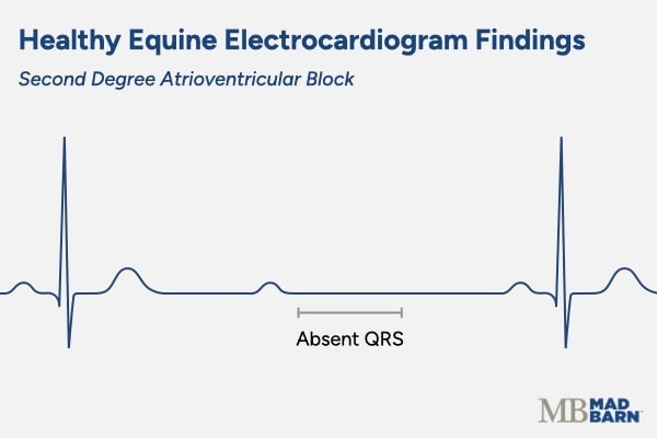 Illustration: Dr. Madison Ricard, DVM, PhD, DACVP, PAS
On the ECG, the P wave is present due to atrial contraction, but the QRS and T waves are not. [2] This arrhythmia is common in horses, particularly very physically fit high-performance horses, and disappears during exercise. [3]
Sinus Arrhythmia
Sinus arrhythmia describes a heart rate that slows down and speeds up alongside the horse’s breathing at rest. [3] This is a normal finding in animals recovering from exercise and some very fit animals at rest. The trigger is high vagal tone caused by exercise.
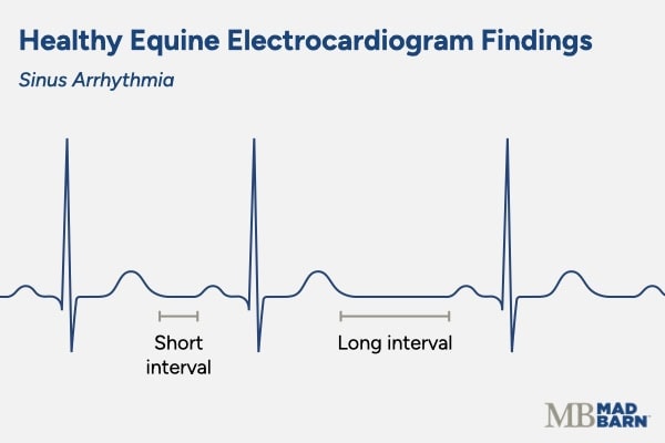 Illustration: Dr. Madison Ricard, DVM, PhD, DACVP, PAS
When the horse breathes out, activation of the vagal nerve causes the heart rate to slow significantly. During inhalation, the vagal nerve is inhibited, increasing heart rate back to normal levels. [3]
Sinus Block
Sinus block is similar to sinus arrhythmia, but activation of the vagal nerve causes a complete stop in heartbeat production. This produces a straight line on the ECG with no waves.
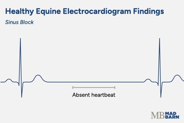 Illustration: Dr. Madison Ricard, DVM, PhD, DACVP, PAS
This is a normal finding in some resting horses that should disappear with exercise. [4] If these “blocks” where no heartbeats are produced are frequent or do not disappear with exercise, then the arrhythmia is pathogenic (disease-causing).
Pathogenic Arrhythmias
Pathogenic arrhythmias are disease-causing, and may result in clinical signs such as poor performance, exercise intolerance, weakness, or collapse. [2]
Atrial Fibrillation
Atrial fibrillation is a very common arrhythmia in horses, with some studies showing that up to 0.3% of racehorses have this arrhythmia. [2] On an ECG, atrial fibrillation shows no discernable P waves, which are replaced with irregular F waves. [2]
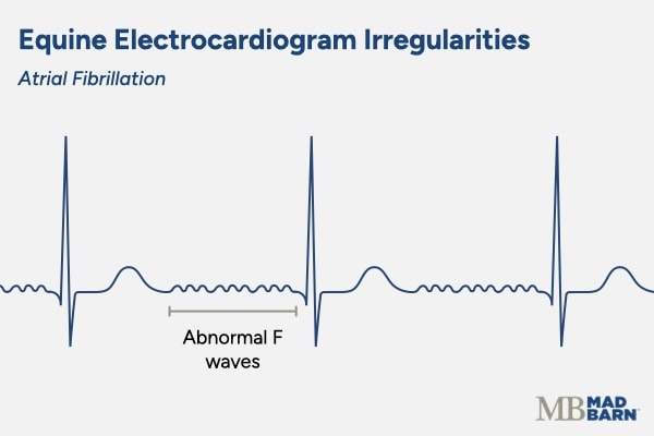 Illustration: Dr. Madison Ricard, DVM, PhD, DACVP, PAS
Common clinical signs associated with atrial fibrillation in horses include: [2]
- Rapid breathing or difficulty breathing
- Poor performance
- Colic-like symptoms
- Coughing
Many cases of atrial fibrillation in horses are considered idiopathic with no identifiable cause or risk factor [5]. With that being said, there are several risk factors, including: [2][5]
- Electrolyte imbalances
- Cardiac disease, such as mitral valve regurgitation
- Use of certain medications, such as furosemide
- High intensity exercise
- Anesthesia
Advanced Atrioventricular Blocks
Some second degree atrioventricular blocks are pathologic, with the horse showing signs of severe exercise intolerance and collapse. [2] On ECG, these horses have multiple skipped beats (no QRS or T complexes) in a row. [2]
In a complete, third-degree atrioventricular block, there are no “normal” ECG waves. Instead, the P waves and QRS complexes are completely disunited.
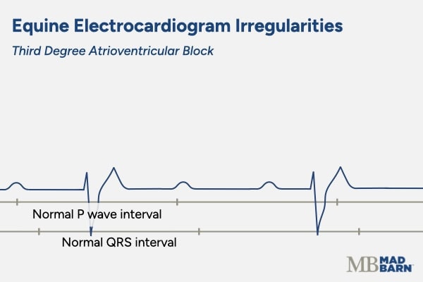 Illustration: Dr. Madison Ricard, DVM, PhD, DACVP, PAS
This occurs when the atrioventricular node does not respond to signalling from the sinoatrial node, so it does not stimulate ventricular contraction. [2] The ventricles can stimulate their own contraction if they do not receive adequate signalling from the atrioventricular node. These contractions are called escape complexes and do not follow a P wave. [2]
Causes of this condition include electrolyte imbalances, digitalis toxicity, and inflammatory or degenerative diseases of the atrioventricular node. [2]
Supraventricular Premature Depolarizations
In this condition, the atria contract on their own without stimulation by the sinoatrial node. [2] On ECG, there are bizarre P waves followed by a normal QRS complex.
 Illustration: Dr. Madison Ricard, DVM, PhD, DACVP, PAS
This condition typically results from heart disease, such as degeneration of the atrioventricular valves or congenital heart disorders. [2]
Ventricular Premature Depolarizations
This condition occurs when the ventricles contract on their own, without stimulation by the atrioventricular node. [2] On ECG, they look like abnormal QRS complexes that have no preceding P wave. [2]
 Illustration: Dr. Madison Ricard, DVM, PhD, DACVP, PAS
If more than four of these complexes occur in a row, it is called ventricular tachycardia.
Occasional premature ventricular depolarizations can occur in resting horses, as well as during and following exercise. [2] However, if they are frequent or have an unusual shape on the ECG tracing, they are considered abnormal.
Causes of ventricular premature depolarizations include inflammation of the heart, electrolyte abnormalities, low blood oxygen, or endotoxemia. [2] They are also common in horses with heart failure. [2]
Complications
ECGs are a non-invasive procedure with no associated complications for the horse.
In some cases, excessive movement of the horse or inadequate contact between the electrodes and the skin may lead to a poor-quality recording. [1]
When this happens, the veterinarian corrects whatever problem is affecting the recording and repeats the procedure until a quality tracing is produced.
Frequently Asked Questions
Here are some frequently asked questions about equine electrocardiography (ECG):
An electrocardiogram (ECG, also known as EKG) is a diagnostic test that records the electrical activity of a horse's heart. It helps veterinarians evaluate heart rhythm and detect abnormalities
Your veterinarian may recommend an ECG if your horse has an irregular heartbeat, poor performance, weakness episodes, or before undergoing anesthesia. It is a valuable tool for diagnosing arrhythmias and other cardiac issues
Electrodes are placed on the horse's skin, usually on the chest and neck areas, to pick up the heart's electrical signals. The procedure is non-invasive, painless, and can often be done in the barn or clinic
No, an ECG does not detect all equine heart problems. While ECGs are excellent for detecting rhythm abnormalities, they do not provide detailed information about heart structure or blood flow. Other tests, such as echocardiography, may be needed for a complete assessment
Yes, ECG is safe for horses. The procedure is non-invasive, painless, and poses no risk of complications
A standard ECG recording usually takes only a few minutes. In some cases, your veterinarian may recommend longer monitoring (Holter ECG) to catch intermittent rhythm abnormalities.
Summary
Electrocardiograms (ECGs) are a key tool for diagnosing and monitoring heart rhythm abnormalities in horses.
- An ECG records the heart's electrical activity to detect arrhythmias (irregular heartbeats)
- The ECG procedure is non-invasive and painless, with no associated complications
- Commonly recommended for horses with irregular heartbeats, weakness or collapse, or poor performance
- Can be performed quickly in a barn or clinic, with results available immediately or via longer monitoring if required
References
- Mitchell. K. J. Equine Electrocardiography. Veterinary Clinics of North America: Equine Practice. 2019.
- Marr. C. M. and Bowen. I. M. Eds. Cardiology of the Horse. 2nd ed. Saunders, Edinburgh; New York. 2010.
- Menzies-Gow. N. ECG Interpretation in the Horse. In Practice. 2001.
- Barton. M. A Guide to Differential Diagnosis of Arrhythmias in Horses. DVM 360. 2008.
- Decloedt. A. et al. Atrial Fibrillation in Horses Part 1: Pathophysiology. The Veterinary Journal. 2020. View Summary


