


Rectal examination remains one of the most valuable and informative diagnostic tools available to equine practitioners evaluating gastrointestinal conditions. In cases of colic, it is often the single most informative procedure for identifying the underlying cause, determining the severity of disease, and guiding immediate treatment decisions.
During a rectal examination, the veterinarian systematically evaluates the accessible organs within the caudal abdomen and pelvis, including the large and small colon, cecum, and bladder. Palpation allows detection of abnormalities that may indicate specific conditions such as large colon volvulus, cecal impaction, or small intestinal obstruction.
Although routine for experienced clinicians, rectal examination must be performed with care to ensure safety for both horse and examiner. Proper restraint, adequate lubrication, and the use of sedation or smooth muscle relaxants are essential to reduce straining and minimize the risk of rectal tears, a serious potential complication.
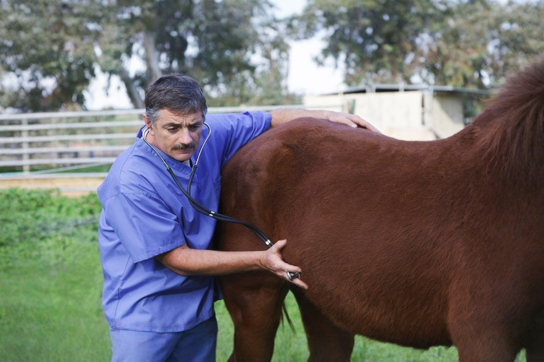
Rectal findings contribute to a comprehensive assessment of gastrointestinal function when combined with other diagnostic approaches, such as abdominal auscultation (listening with a stethoscope), nasogastric intubation, ultrasonography, and laboratory testing. For veterinarians managing horses with colic or other digestive disturbances, mastery of this procedure and the ability to interpret subtle variations in normal anatomy are critical components of effective case management.
Rectal Examinations in Horses
Rectal examination is a key component of the diagnostic workup for equine colic and other gastrointestinal disorders.
Veterinarians use this procedure to assess the location and integrity of the large intestine and other abdominal organs. This can help them determine whether a horse requires surgical intervention to resolve their colic signs, or if they can be managed using medications alone.
Common signs of colic in horses indicating the need for a rectal examination include: [1]
- Lack of appetite
- Lethargy
- Looking at the flanks
- Pawing
- Stretching out as if to urinate
- Kicking at the abdomen
- Restlessness
- Rolling or thrashing
- Unwilling to stand
Other signs of gastrointestinal disease that may lead to a rectal examination include: [1]
- Straining to defecate
- Weight loss for unknown reasons
- Chronic, mild colic signs or inappetence

Equipment
Rectal examination requires minimal equipment. The veterinarian dons a shoulder-length plastic palpation sleeve and applies a large amount of lubricant to the glove surface. The lubricant helps reduce the risk of damage to the delicate mucosa lining the horse’s rectum. [1]
Most veterinarians prefer to perform rectal examination in a set of stocks. [1] This helps protect the veterinarian if the horse kicks backwards during the procedure.
Alternatively, they may use a stack of square hay bales to make a short barrier. They can also position the horse with their hindquarters facing the door of a stall and stand to the side of the horse using the stall wall for protection. [1]
Many veterinarians also place a twitch on the horse’s muzzle to provide a distraction. [2]
What's your top priority with your horse's health?
Enter your email to receive your store credit
Procedure
Many horses require sedation for rectal examination, particularly if they are in gastrointestinal distress and showing signs of colic. This helps ensure they stand quietly during the procedure, reducing the risk of injury to the rectal mucosa and the veterinarian. [1]
Before starting the exam, the veterinarian administers a sedative to the horse and allows 5 – 10 minutes for the medication to take effect.
The veterinarian may also administer N-butylscopolammonium bromide, a medication that prevents spasm (contraction) of the rectal tissue. This helps prevent the horse from straining against the veterinarian’s hand, allowing for a smoother procedure and a lower risk of rectal tears. [1] Alternatively, they may give a lidocaine enema to achieve a similar effect.
Once the horse is sedated, the veterinarian positions themselves to the side of the horse’s hindquarters. They slowly reach under the tail and insert their gloved handed into the horse’s anus. [1] As the horse relaxes, they advance their hand into the rectum.
The horse’s rectum is typically filled with manure that must be removed before the veterinarian can begin their examination. The veterinarian carefully removes the feces by gently grasping the fecal balls and scooping them out of the rectum. [1] It may take several attempts to remove all of the feces from the rectum. [1]
After emptying the rectum, the examination begins. The veterinarian palpates all four quadrants of the abdomen, as well as the dorsal (towards the spine) and ventral (towards the ground) midlines. [1] They palpate the structures present in each quadrant to ensure they are the expected size, in the correct location, and have no notable abnormalities.
Once the examination is complete, the veterinarian withdraws their hand and inspects their glove for blood. Significant blood on the palpation sleeve may indicate that a rectal tear occurred, which requires immediate surgical intervention.
Interpreting Results
As the veterinarian palpates each quadrant, they feel for specific structures. Changes in these structures may indicate the underlying cause of the horse’s abdominal pain and colic signs.
Most veterinarians start by palpating one quadrant, then work in a clockwise direction. [1] Having a consistent approach helps ensure nothing is missed during the examination.
Although the rectal examination can provide a wealth of information about the horse’s abdomen, several structures are out of reach for the veterinarian. Estimates suggest that only 30 – 40% of the horse’s abdomen can be examined through rectal palpation. [2]
Structures that cannot be examined include: [3]
- Stomach
- Right kidney
- Liver
- Transverse colon
- Cranial (towards the head) portions of the large colon
Some structures are only palpable if they are in an abnormal location, including: [3]
- Small intestine
- Mesenteric root
- Right dorsal colon
Left Dorsal Quadrant
In the left dorsal quadrant, the veterinarian should be able to feel the horse’s left kidney and the edge of the spleen. [1] The spleen should be against the body wall in a healthy horse with no abnormalities. [3]
In some cases, the veterinarian may feel intestinal loops between the spleen and body wall, a condition called nephrosplenic ligament entrapment. [3]
Dorsal Midline
Feeling upwards towards the horse’s spine, the veterinarian can palpate the horse’s aorta, the largest artery in the body. They can also feel the iliac arteries, large arteries that supply the hind limbs.
The pulse in the left and right iliac arteries should be similar. Differences in the pulse intensity may indicate a thrombus (blockage) that can cause severe pain and may be life-limiting. [3]
Right Dorsal & Ventral Quadrants
The right dorsal quadrant contains the head of the cecum. The cecum is a large comma-shaped extension of the horse’s large intestine that plays a major role in fiber fermentation. In a normal horse, the cecum is typically empty or contains only a small amount of feed material and gas. [3]
A gas-filled cecum is called cecal tympany, while a feed-filled cecum is a cecal impaction. [3] In some cases, the veterinarian may feel a mass near the cecum. This may be an intussusception, a condition where the intestine telescopes over itself, preventing passage of feed. [3]
The right ventral quadrant contains the body of the cecum. The tail of the cecum is usually not palpable, as it sits out of reach against the horse’s ventral abdominal wall. The veterinarian continues to palpate the cecum and ensures there are no unexpected structures in the area. [3]
Ventral Midline
Along the ventral midline, the veterinarian can palpate the horse’s pelvis. Identifying unusual ridges or changes can indicate a pelvic fracture. The bladder may be palpable if it is filled with urine. [3]
In mares, the uterus and ovaries are palpable from their position resting within the pelvis. [3] Rectal examinations are also an integral part of breeding evaluations in mares. The veterinarian can palpate the uterus, both ovaries, and may be able to palpate a fetus in a pregnant mare. [3]
In male horses, especially stallions, the veterinarian can palpate the inguinal rings, gaps in the abdominal wall that the spermatic cord passes through. [3] Segments of intestine can slip into these rings causing an inguinal hernia, a painful and uncomfortable condition that often requires surgical intervention.
As the veterinarian examines the pelvic region, they may feel a section of large colon running from right to left. This indicates a right dorsal displacement, where the large colon has rotated within the abdomen. Right dorsal displacement typically requires surgical intervention to correct. [3]
Left Ventral Quadrant
The left ventral quadrant contains the pelvic flexure, a 180º degree turn in the colon. This is the most common location where impactions develop, as the colon narrows dramatically prior to the flexure, preventing passage of bulky feed materials. [1]
The small descending colon also runs through the left ventral quadrant, allowing the veterinarian to assess fecal ball production. [3]
Small Intestine
The small intestine may be palpable in any quadrant when there is small intestinal disease present. It is not normally palpable in a healthy horse.
A dilated loop of small intestine filled with fluid typically indicates there is a blockage preventing normal passage of feed. A loop of small intestine with a thick wall usually indicates infection or inflammation.
Potential causes of a palpable small intestine include: [3]
- Hernias
- Intussusception
- Ileal impaction
- Strangulating lipoma
- Functional stasis of the intestinal tract (ileus)
- Proliferative enteropathy
- Proximal enteritis

Preparing Your Horse for Rectal Examination
There are no specific steps required to prepare your horse for a rectal examination. If possible, have your horse located in an area with stocks or a nearby stall that the veterinarian can use for restraint.
Your veterinarian may have you haul to their clinic to ensure adequate restraint facilities are available.
After the procedure, your horse must wake up from sedation before they are allowed to eat. Having a stall or small paddock with no accessible feed is ideal.
It can take up to an hour for the horse to recover from sedation and return to its normal living areas.
Complications
The most common complication associated with rectal examination is a rectal tear. [3] These tears occur from the veterinarian’s hand causing irritation or injury to the delicate lining of the rectum.
Preventative measures that veterinarians employ to prevent rectal tears include: [3]
- Appropriate restraint, including sedation if necessary
- Ample lubrication
- Use of medications to prevent contraction of the rectum
- Removing their hand when contractions occur
- Keeping fingernails short to avoid snagging or damage
- Using specialized equine palpation sleeves with little to no seam
Rectal tears are an emergency and require rapid intervention to prevent fecal material from leaking into the horse’s abdomen. Careful palpation allows the veterinarian to determine how deep the tear is, as well as the length of the tear and degree of fecal contamination. [3]
The depth of the tear correlates with the horse’s prognosis and its treatment: [3][4][5]
- Grade 1: These tears only affect the mucosa and submucosa. They have an excellent prognosis when treated with antibiotics and anti-inflammatories. Horses are also put on soft, lubricating feed like fresh grass to reduce straining during defecation.
- Grade 2: These tears affect the muscle layer. They have a similar prognosis and treatment to Grade 1 tears.
- Grade 3: All layers are affected except the serosa, a thin layer of connective tissue surrounding the rectum. These horses require immediate surgery to repair the rectum. The prognosis for survival is around 35%.
- Grade 4: Complete tear through the rectal wall. Even with immediate surgical repair, the prognosis for survival is around 2–6% for these horses.
In the case of Grade 3 or 4 tears, the veterinarian must intervene immediately to help prevent fecal material from entering the abdomen. Emergency first aid involves packing the rectum with lubricated cotton to prevent passage of feces.
This procedure is typically combined with an epidural, to prevent the horse from straining against the pack and potentially worsening the tear. [5]
Frequently Asked Questions
Here are some frequently asked questions about rectal examination in horses:
Rectal examination is a critical diagnostic procedure used to assess the caudal abdomen and pelvis of the horse. It allows the veterinarian to palpate structures such as the large colon, small colon, cecum, bladder, and reproductive organs. In cases of suspected gastrointestinal disease, like colic, it provides valuable information about intestinal position, distension, and content consistency.
During an equine rectal examination, veterinarians may identify gas or fluid distension of the intestines, displacement or impaction of the large colon, cecal distension, small intestinal loops, or abnormalities in the small colon. These findings can provide clues to specific conditions like large colon volvulus, cecal impaction, or small intestinal obstruction.
While generally safe in skilled hands, rectal tears are a recognized risk of rectal examination, and can be life-threatening. Gentle technique, adequate lubrication, and proper restraint are essential to minimize trauma. Sedation and the use of antispasmodic medication can reduce straining and improve safety.
While rectal findings can strongly suggest specific gastrointestinal disorders, they are best interpreted alongside clinical signs, nasogastric intubation results, abdominal ultrasound, and laboratory data. Together, these tests allow for accurate diagnosis and treatment planning.
Summary
Rectal examination is a diagnostic tool for assessing gastrointestinal disorders in horses, offering direct insight into the abdominal organs and helping guide emergency and medical management.
- Rectal examination allows palpation of large colon, cecum, small colon, bladder, and reproductive structures
- It can be used to identify abnormalities such as impactions, displacements, and intestinal distension
- The examination must be performed with careful restraint, sedation, and lubrication to minimize risk of rectal tears
- Results of rectal examinations in horses should be interpreted in conjunction with other diagnostic findings for accurate assessment
References
- Leith. G. et al. Eds. Performing the Large Animal Physical Examination. 1st ed. Wiley. 2025.
- Blikslager. A. T. et al., Eds. The Equine Acute Abdomen. Third edition. Wiley, Blackwell, Hoboken, NJ. 2017.
- Southwood. L. L., Ed. Practical Guide to Equine Colic. Wiley-Blackwell, Ames, Iowa. 2013.
- Costa. L. R. R. and Paradis. M. R. Manual of Clinical Procedures in the Horse. Wiley Blackwell, Hoboken. 2018.
- McMaster. M. et al. A Review of Equine Rectal Tears and Current Methods of Treatment. Equine Veterinary Education. 2015.


