


Nasogastric intubation is one of the most frequently performed procedures in equine practice. It serves both diagnostic and therapeutic purposes and is critical for evaluating horses with gastrointestinal disease, particularly those presenting with signs of colic.
Although the procedure might look uncomfortable, when performed correctly by a trained professional, it is safe and provides crucial information about what is happening inside a horse’s digestive system.
Because horses are unable to vomit, the ability to safely decompress the stomach and assess gastric contents is lifesaving in certain situations. Beyond its emergency applications, nasogastric intubation is also used for medication administration, nutritional support, and gastric lavage.
Proper technique is essential to avoid injuring the horse or placing the tube in the wrong location. For this reason, nasogastric intubation should always be performed by a veterinarian or under their direct supervision to ensure it is safe and effective. This article discusses the process of nasogastric intubation and potential complications of the procedure.
Nasogastric Intubation in Horses
Nasogastric intubation is a critical component of a routine colic exam. Not only does this procedure help veterinarians diagnose the potential cause of colic signs, but it is also an important component of treatment.
Veterinarians combine the results of nasogastric intubation with rectal and physical examination findings to determine whether a horse requires surgical intervention or can be managed with medication alone.
Signs of colic in horses include: [1]
- Lack of appetite
- Lethargy
- Looking at the flanks
- Pawing
- Stretching out as if to urinate
- Kicking at the abdomen
- Restlessness
- Rolling or thrashing
- Unwilling to stand
Depending on the horse’s physical examination findings, the veterinarian may perform nasogastric intubation before rectal examination. This is usually done in cases where the horse has a high heart rate, which can indicate there is reflux of digesta from the small intestine into the stomach, causing painful and life-threatening distension. [2]
Passing the tube in these cases helps relieve the fluid accumulation and makes the horse more comfortable during further diagnostic testing. [2]
What's your top priority with your horse's health?
Enter your email to receive your store credit
Equipment
Nasogastric intubation requires very little equipment. The main piece of equipment is the nasogastric tube, a long flexible plastic tube with a smooth surface. [2] The size and length of the tube used depends on the size of the horse and the veterinarian’s preference. [2]
Other equipment includes buckets of water and a stomach pump. The veterinarian can attach the end of the nasogastric tube to the stomach pump to form a siphon that removes fluid from the stomach. They can also use the pump to deliver treatments, such as fluids and medication.
Procedure
Horses often require sedation for nasogastric intubation. The procedure itself isn’t painful, but horses may be uncomfortable and difficult to handle because of their colic. Sedation also helps keep the veterinarian safe by reducing the risk of the horse striking out or rearing. [2]
Some horses may also require a nose twitch to provide a distraction as the tube is passed. [2]
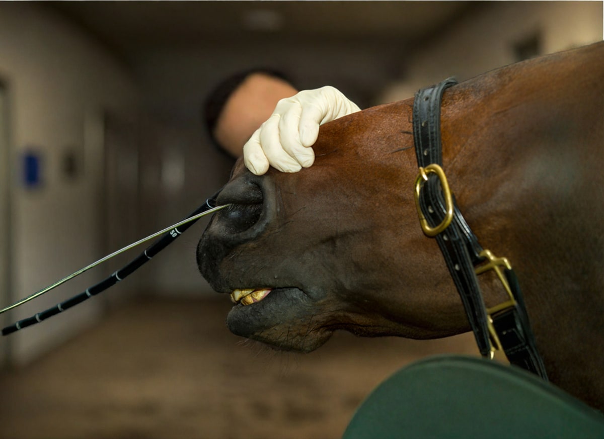
Once the horse is sedated, the veterinarian can begin the procedure. They lubricate the tip of the nasogastric tube with mineral oil or water to reduce the risk of damaging the delicate nasal tissues. [2] They introduce the tube into the horse’s nostril and guide it carefully through the nasal passages to the pharynx (throat). [2]
They may flex or extend the horse’s head to try and aim the nasogastric tube towards the horse’s esophagus, rather than into the trachea. In response to the tube’s presence in the pharynx, the horse may attempt to swallow. This movement helps the veterinarian guide the tube into the horse’s esophagus. [2]
Once the veterinarian has placed the tube, they need to confirm it is in the esophagus and not the trachea. Delivery of fluids into the lungs can be fatal. To check that placement is correct, they observe the tip of the tube moving down the horse’s neck. [2] They may also wiggle the horse’s trachea to feel for rattling, indicating that the tube is within the trachea.
If the tube is in the esophagus, the veterinarian proceeds to pass the tube further into the horse’s digestive system to the stomach. Upon entry to the stomach, the tube often releases odorous gas that smells like acidic or fermenting feed.
The veterinarian may blow in the tube while their assistant listens to the horse’s abdomen with a stethoscope. Blowing in the tube typically produces a bubbling or gurgling sound that indicates correct placement. [2]
Once the tube is in position, the veterinarian can assess for reflux (excess fluid within the stomach). They attach the end of the tube to the stomach pump and deliver around 1 L of water through the tube. [2] Once the tube is “primed” and has formed a siphon, they remove the stomach pump and allow fluid to drain out from the tube into a bucket. [2]
The horse’s stomach capacity is typically around 8 – 15 L. When there is excess fluid, it may contain up to 20 L, which is a dangerous amount that can result in gastric rupture. [2] As the fluid drains from the horse’s stomach, the veterinarian measures the quantity of fluid to determine whether there is net reflux. They may repeat the siphoning procedure several times to ensure the stomach is completely empty before determining their final measurement. [2]
Depending on the results, the veterinarian may leave the nasogastric tube in place for future use, administer fluids directly into the stomach, or they may remove the tube. [2] To remove the tube, they kink the tube to prevent fluid from escaping, then gently pull the tube out of the horse’s nostril. [2]
Interpreting Results
Nasogastric intubation provides the veterinarian with information about the type of colic occurring and if there’s fluid from the small intestine backing up into the stomach (reflux). They can evaluate the amount of fluid returned from the procedure, as well as its color and consistency. They may also submit a sample of reflux for bacterial testing. [2]
Less than 2 L of net reflux from the stomach is considered within normal limits. Horses that produce >2 L net reflux likely have a blockage preventing fluid in the stomach or small intestine from passing into the large colon. [2] They may also have an inflammatory condition of the small intestine causing excess fluid production, resulting in fluid backing up into the stomach.
Possible causes of reflux include: [2]
- Ileus
- Proximal enteritis
- Small intestinal obstruction, such as from intussusception or strangulating lipomas
The color can also indicate the type of fluid present. Healthy stomach fluid is green or brown, while yellow can indicate reflux from the small intestine. [2] Orange or red may indicate there is hemorrhagic (bloody) enteritis within the small intestine. [2]
Finally, the veterinarian can also assess the ease of passing the tube. Gastric impactions, accumulations of feed material within the stomach, can make passing a nasogastric tube difficult. [2] They can also prevent siphoning of fluid by blocking the tip of the tube.
Preparing Your Horse for Nasogastric Intubation
There are no specific preparations required for nasogastric intubation. Before the veterinarian arrives, position your horse in a well-lit area with a slip-resistant floor. If available, having your horse in restraint facilities such as stocks is ideal.
Since most nasogastric intubations are performed as part of a colic work-up, it is important to know that your horse will be put on a restricted diet after the procedure.
Most veterinarians recommend keeping the horse off feed for at least 24 hours, before slowly reintroducing hay.
Have a grass-free paddock or stall prepared for your horse to restrict their feed intake during their recovery process.

Complications
Overall, nasogastric intubation is a very safe procedure when performed by an experienced veterinarian. However, There are several potential complications of nasogastric intubation that are more likely to occur when non-veterinarians or owners attempt the procedure.
Potential complications include: [3]
- Nosebleeds
- Trauma or irritation of the esophagus or pharynx
- Tube breaking and subsequent ingestion
- Fluid administration into the lungs
- Sinusitis
Nosebleeds
The most common complication associated with nasogastric intubation is bleeding from the nose. [3] Bleeding either originates from the sensitive nasal lining or the ethmoid turbinates.
The ethmoid turbinates are a collection of scroll-shaped bones at the back of the horse’s nasal sinuses. These bones are covered by highly vascularized, delicate tissue that bleeds easily when touched by a nasogastric tube. [3]
In most cases, the bleeding stops on its own and the horse recovers uneventfully. Rarely, bleeding can be severe enough to require a blood transfusion and drugs to promote blood coagulation. [3]
To reduce the risk of nosebleeds, veterinarians are trained to pass the tube in a manner that follows the horse’s anatomy using a gentle technique. [3]
Additionally, they factor in the patient’s size when selecting their tube, to reduce the chances of interacting with the ethmoids. Adequate sedation and restraint by an experienced handler can also help reduce the risk of bleeding.
Esophageal & Pharyngeal Trauma
Passing a tube through the pharynx and esophagus can cause local irritation to the tissues. [3] This can result in ulcerations, tears, and potentially perforation of the tissue at any point along the horse’s neck. [3][4]
Horses that resist intubation by retching and contracting their neck muscles have a higher risk of traumatic injury. Smaller horses or horses who have nasogastric tubes left in for long periods also have a higher risk. [3]
Signs of traumatic injuries in the esophagus and pharynx include: [3][5]
- Coughing
- Difficulty swallowing
- Excessive salivation
- Grinding teeth
- Fever
- Swelling in the neck
Treatment typically involves anti-inflammatory medications and soft or soupy feed to prevent additional damage to the tissue. [3] Some horses may also require antibiotics to treat local infections. Rarely, surgical intervention to remove abscesses or infected tissues is necessary. [3]
The overall prognosis for survival is good, but horses may develop scarring in the esophagus that increases their risk of choke later in life. [3]
Tube Breaking
Rarely, the nasogastric tube may break due to structural failure of the plastic. [3] This can occur from repeated use of the tube or exposure to damaging environmental conditions such as sunlight or cold temperatures.
Horses can also bite the tube and break it. This occurs when the tube inadvertently bends backwards into the horse’s oral cavity, something that is more likely to happen when non-veterinarians attempt to intubate.
Veterinarians routinely inspect their nasogastric tubes for signs of damage or brittleness as a preventive measure. [3] When horses swallow a piece of tube, treatment requires removal of the tube fragments using an endoscope or surgical intervention. [3]
Fluid in the Lungs
One of the most severe complications associated with nasogastric intubation is the delivery of fluids into the lungs from a misplaced tube. [3] For this reason, veterinarians check the tube’s placement several times before passing any fluid.
The severity of this complication depends on the amount and type of fluids administered. If only a small amount of clean water is administered, treatment typically involves anti-inflammatory medications and antibiotics to prevent pneumonia. [3] Large amounts of clean water may require additional supportive care such as furosemide to reduce fluid in the lungs and oxygen therapy. [3]
The most severe consequences occur when medications or mineral oil are pumped into the lungs. These incidents are nearly always fatal as the lungs cannot remove the foreign material, resulting in a severe pneumonia and tissue death. [3]
Sinusitis
Sinusitis is an infection in the nasal passages. It is most commonly associated with prolonged nasogastric intubation or repeated intubations. [3] Horses with sinusitis develop fever and profuse cloudy or pus-like nasal discharge.
Treatment involves antibiotics and lavage (flushing) of the sinuses to remove the infection. Most horses recover fully with appropriate treatment. [3]
Frequently Asked Questions
Here are some frequently asked questions about nasogastric intubation in horses:
Nasogastric intubation is a veterinary procedure in which a flexible tube is passed through a horse's nostril, down the esophagus, and into the stomach to assess gastric contents, relieve pressure, or administer fluids and medications.
It is most often performed in horses showing signs of colic to evaluate stomach contents, relieve fluid buildup, or administer treatments such as fluids, mineral oil, electrolytes, or medications.
The quantity, color, and odor of refluxed fluid provide diagnostic clues about the horse's gastrointestinal function. Large or abnormal reflux may indicate obstruction or inflammation.
Possible complications of nasogastric intubation for horses include nosebleeds, nasal tissue trauma, esophageal injury, accidental tracheal placement, or aspiration of fluids. Proper restraint and technique minimize these risks.
No, owners should never try to perform nasogastric intubation for horses on their own. This procedure must only be performed by a veterinarian or under veterinary supervision to prevent injury or complications.
Summary
Nasogastric intubation is a vital diagnostic and therapeutic procedure in equine medicine, primarily used to assess and relieve gastric distension in horses with colic.
- The procedure involves passing a well-lubricated tube through one of the horse’s nostrils and into the stomach.
- It is an essential technique for evaluating gastric reflux and for administering fluids or medications directly into the stomach.
- The results can provide important diagnostic information about gastrointestinal function and potential obstruction.
- This procedure should only be performed by a trained veterinarian to minimize the risk of injury or other complications.
References
- Leith. G. et al., Eds. Performing the Large Animal Physical Examination. 1st ed. Wiley. 2025.
- Southwood. L. L., Ed. Practical Guide to Equine Colic. Wiley-Blackwell, Ames, Iowa. 2013.
- Rubio-Martinez. L. M. and Hendrickson. D. A. Complications in Equine Surgery. Wiley Blackwell, Hoboken. 2021.
- Byrne. C. A., Challenges and Complications of Prolonged Nasogastric Intubation in Equine Patients. Equine Veterinary Education. 2025.
- Hardy. J. et al., Complications of Nasogastric Intubation in Horses: Nine Cases (1987–1989). Journal of the American Veterinary Medical Association. American Veterinary Medical Association. 1992.


