


The nervous system is involved in every aspect of a horse’s health and performance, from balance and coordination to sensory perception and behavior. When this system is interrupted, the signs can be subtle or dramatic, and distinguishing neurologic disease from other causes of poor performance or lameness can be challenging.
A neurologic examination is a systematic process that veterinarians use to evaluate the function of the brain, spinal cord, and peripheral nerves in horses.
By carefully observing the horse at rest and in motion, and by performing specific tests of reflexes, coordination, and responses, veterinarians can identify abnormalities and localize problems within the nervous system.
For horse owners, understanding what to expect during a neurologic exam can help reduce stress and highlight the importance of early veterinary intervention. Detecting neurologic problems promptly not only improves outcomes but also plays a critical role in ensuring the safety of both horse and handler.
Equine Neurologic Examinations
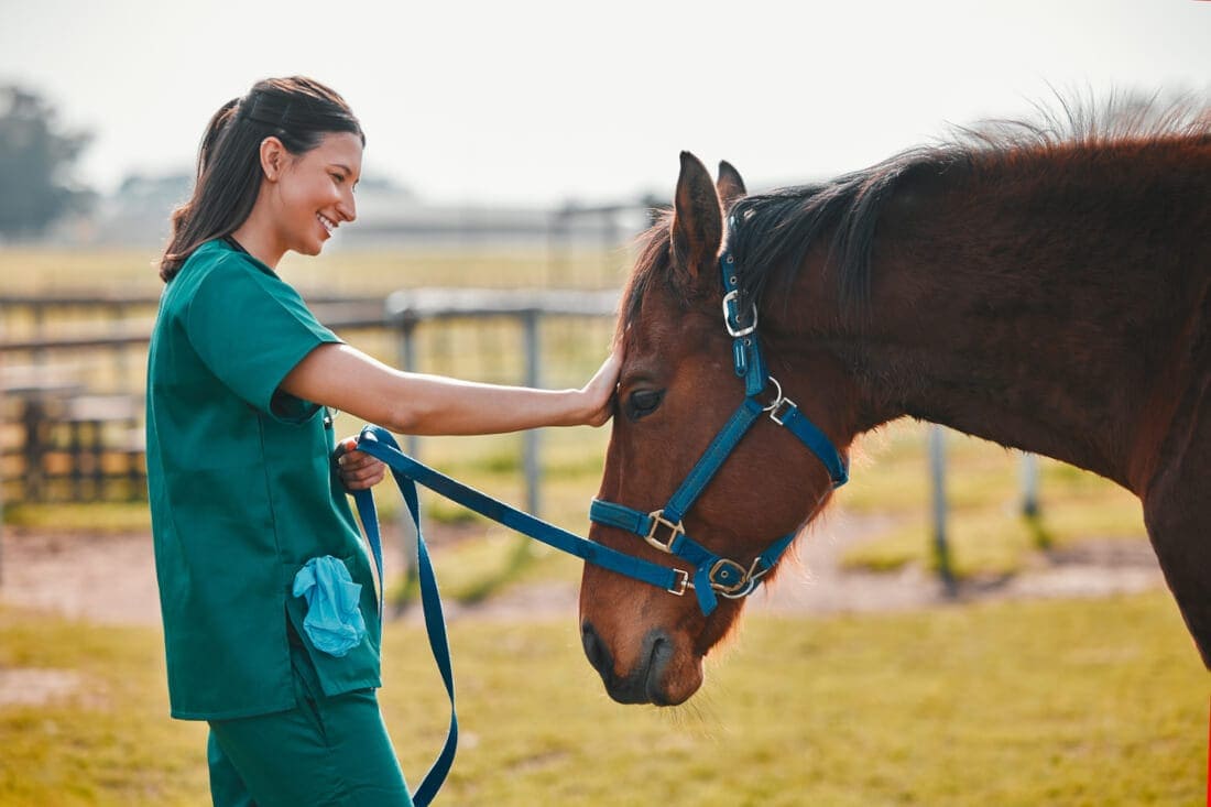
Veterinarians use the neurologic examination to determine if there are any abnormalities in the horse’s nervous system. By performing a thorough examination, they can assess areas of the horse’s brain, spinal cord, or nerves that may be diseased.
Signs of neurologic disease in horses vary widely, depending on which part of the nervous system is affected. Clinical signs may include: [1]
- Uncoordinated movement (ataxia)
- Stumbling or tripping
- Exaggerated limb movement
- Head tremors
- Extreme lethargy
- Weakness
- Paralysis
- Seizures
- Blindness
- Asymmetry of the face
- Inability to blink
- Inability to swallow
- Flaccid tongue
- Urinary incontinence
- Changes in attitude or mentation
- Muscle atrophy
Equipment
Minimal equipment is required for a neurologic examination. Equipment typically includes: [2]
- A pen light or flashlight
- Hemostats or a pen for poking the skin
- A towel to use as a blindfold
- A halter and leadshank to restrain the horse
An assistant in charge of restraining and leading the horse is also necessary.
What's your top priority with your horse's health?
Enter your email to receive your store credit
Procedure
The general procedure for a neurologic examination can vary between veterinarians, but the main goals are assessing the horse’s overall behavior and condition, their cranial nerve function, and their gait and posture.
General Examination
The veterinarian begins their examination by observing the horse’s resting behavior. This allows them to identify signs of lethargy or proprioceptive deficits, meaning the horse is having difficulty with orientation within the environment. They can also observe any unusual postures or stances the horse performs. [2]
Closer inspection of the symmetry of the horse allows the veterinarian to identify muscle atrophy (loss), which can indicate that a nerve is not working correctly. [2]
Using a pen or other pointy object, the veterinarian may poke different areas of the horse’s skin to see if there is a reaction.
The horse should twitch their skin or move away from the pressure. Failing to do so indicates that there is a nerve deficit in the nerves supplying that area of skin or there is pain preventing the horse from moving away from pressure. [2][3]
They also manipulate the horse’s tail to ensure there is adequate muscle tone in that region of the body.
Cranial Nerve Examination
The cranial nerves are a series of nerves emerging directly from the brain that provide sensation, movement, and activation functions to structures of the head and neck.
Identifying deficits in the cranial nerves can indicate a problem within the nerve itself, or a problem within the brain resulting in impaired nerve function. [2]
There are twelve cranial nerves, and veterinarians test all of their functions during the neurologic examination. [2]
During the cranial nerve examination, the veterinarian performs a series of reflex tests, including the menace response and a blindfolded test of smelling, hearing and balance, to assess the horse’s cranial nerves.
Olfactory Nerve
The olfactory nerve is responsible for the horse’s sense of smell. Offering feed or an unusual smell to a blindfolded horse and identifying their reaction can help detect problems in this nerve. [2]
Optic Nerve
The optic nerve is responsible for vision. Veterinarians use a menace test to determine if the horse is blind. In this test, they move their hand quickly towards the horse’s eye, and check to see if the horse blinks or moves their head away from the hand. [2]
Oculomotor Nerve
The oculomotor nerve controls the diameter of the eye’s pupil. Shining a bright light into the eye should cause the pupil to constrict if the oculomotor nerve is intact. [2]
Oculomotor, Trochlear, & Abducens Nerves
The oculomotor, trochlear, and abducens nerves control the movement of the eye itself. The veterinarian moves the horse’s head around and watches to see how the eyes respond to that movement. If there are deficits, the eyes either do not respond to the head’s motion, or respond in an unusual fashion. [2]
Trigeminal Nerve
The trigeminal nerve stimulates movement of the chewing muscles and provides sensation to the skin on the face. [2] The veterinarian tests for skin sensitivity by touching the horse’s ears, eyelids, face, and inside of the nostrils to look for a reaction.
They can also test chewing muscle function by offering the horse a treat and looking at their chewing ability. [2]
Facial Nerve
The facial nerve stimulates movement in the facial muscles, allowing the horse to move their ears, eyelids, nostrils, and lips. [2]
Signs of facial nerve deficits in horses include: [2]
- Drooping ear
- Drooping of the lip and nostril
- Reduced tear production by the eye
- Drooping of the eyelid
Vestibulocochlear Nerve
The vestibulocochlear nerve helps with balance and hearing. Applying a blindfold to the horse and observing their movements helps veterinarians identify deficits in this nerve. [2]
They can also test the horse’s response to sound to detect deafness or hearing loss. [2]
Glossopharyngeal, Vagus, & Accessory Nerves
These nerves are responsible for movement of the pharynx and larynx (voice box). Watching the horse swallow can identify issues in the pharynx.
The veterinarian may also perform a “slap test”, where they gently slap the horse on the withers while feeling the horse’s larynx. In a normal horse, the cartilages in the larynx should twitch in response to the slap. [2]
Hypoglossal Nerve
This nerve provides movement to the tongue. The veterinarian pulls the horse’s tongue out of the mouth and watches to see if the horse can retract their tongue on their own. [2]
Gait & Posture
The gait and posture examination involves leading the horse through a series of maneuvers designed to accentuate any movement deficits. [2]
These movements include: [2]
- Walking in a straight line
- Walking with the head raised
- Backing up
- Circling
- Moving up and down hills
Depending on the test, the veterinarian may also blindfold the horse to prevent them from visually compensating for their neurologic deficit. [2] The veterinarian may also pull on the horse’s tail gently while they walk to assess for weakness.
During this evaluation, the veterinarian looks for: [2]
- Weakness, such as dragging the toes or stumbling
- Ataxia (uncoordinated movement)
- Hypermetria causing excessive flexion of the joints which leads to a high-stepping gait
- Spasticity causing decreased flexion of the joints
If the veterinarian identifies a deficit, they grade it on a 5-point scale: [2]
- Grade 0: No deficits noted
- Grade 1: Difficult to see deficits under normal circumstances, but are present when the horse is challenged
- Grade 2: Visible when moving under normal circumstances, such as circling
- Grade 3: Obvious deficits when moving under normal circumstances
- Grade 4: Obvious deficits when standing, and falls or nearly falls during normal movement
- Grade 5: Unable to stand
The veterinarian may also perform limb placement tests to see if the horse understands where its limbs are located (proprioception) and is able to return them to a normal position on their own.
To do this, the veterinarian places the limb in an unusual position, such as: [2]
- Crossing the leg over the opposite limb
- Placing the leg in a very wide stance
- Placing the hoof down on the ground in a flexed position so the horse must move the leg to place the hoof sole back on the ground

Interpreting Results
The results of a neurologic examination help the veterinarian with neuroanatomic localization. This is the process of identifying the location of the horse’s nervous system deficit, so that a list of possible diagnoses can be made. [1]
First, the veterinarian determines whether the cranial nerves are affected. If so, then the condition likely originates in the brain or brainstem. If not, then the condition is affecting the spinal cord. [1]
The Brain
The brain is a complex structure with numerous different components controlling the horse’s body functions. Based on the horse’s clinical signs, the veterinarian can localize the disease to one of three locations. [1]
Cerebrum
The cerebrum is responsible for controlling voluntary movement, processing sensation, and conscious thought. Horses showing signs of seizures, blindness, or delirium likely have cerebral disease.
Cerebral disorders include: [4]
- Hepatic encephalopathy
- Moldy sweet corn toxicosis
- Yellow star thistle toxicosis
- Juvenile epilepsy
- Narcolepsy
- Infectious meningitis or encephalitis
- Trauma
Cerebellum
The cerebellum is responsible for balance and posture. Signs of cerebellar disease include hypermetria (excessive joint flexion while moving), tremors, and weakness.
Conditions affecting the cerebellum include: [4]
- Rabies
- Cerebellar abiotrophy
- Cerebellar hypoplasia
- Trauma
Brainstem
The brainstem regulates automated functions such as breathing and heart rate. Horses with brainstem disease may have altered consciousness, paralysis of all four limbs, or other severe gait deficits.
Example diseases affecting this region include: [4]
The Spinal Cord
The spinal cord runs from the horse’s brainstem to their lumbar spine, where it fans out into several nerve bundles called the cauda equina. One of its primary roles is controlling movement of the limbs.
By grading ataxia (incoordination) in each of the horse’s limbs, veterinarians can determine where spinal cord disease may be located: [1]
- Rear limbs worse than front: If the horse’s rear limbs have a higher ataxia grade than the front limbs, the disease is likely located in the middle to upper neck.
- Front limbs worse: If the front limbs are more severely affected, the region affected is between the lower neck and the front of the withers.
- Only rear limbs affected: If the front limbs are normal but the rear limbs show incoordination, the spinal cord in the horse’s back is likely damaged.
- Tail or bladder paralysis: Horses showing signs of tail or bladder dysfunction likely have damage in their sacrum (lower back) or cauda equina.
There are several conditions affecting the spinal cord of horses, including: [4]
- Cervical vertebral stenotic myelopathy (wobbler syndrome)
- Equine degenerative myeloencephalopathy
- Equine motor neuron disease
- Equine herpesvirus
- Polyneuritis equi
- Equine protozoal myeloencephalitis
- Rabies
- Trauma
The Peripheral Nerves
The peripheral nerves are nerves found throughout the body, outside of the spinal cord. They control muscle movement, sensation, and other functions. Certain diseases may target these nerves, resulting in functional disturbances that a veterinarian can identify during examination.
Examples of peripheral nerve disorders include: [1]
- Stringhalt
- Equine laryngeal paralysis (roars)
- Grass sickness
- Botulism
- Sweeney shoulder
Preparing Your Horse for a Neurologic Examination
There are no specific steps you must take to prepare your horse for a neurologic examination.
Your veterinarian may have you act as the handler during the examination, including trotting and lunging the horse. Have suitable equipment ready, such as a lunge line and whip.
Complications
The neurologic examination is a non-invasive procedure with few complications. Severely uncoordinated horses may fall over during the examination, putting themselves at risk of injury or at risk of injuring their handlers.
Mild stress or resistance may also occur as the horse is asked to perform unfamiliar movements, but these effects are generally temporary.
Frequently Asked Questions
Here are some frequently asked questions about neurologic examinations in horses:
A neurologic examination is a systematic evaluation of a horse's nervous system. It allows veterinarians to assess brain, spinal cord, and peripheral nerve function to detect abnormalities such as weakness, incoordination (ataxia), or abnormal reflexes.
Horses may require a neurologic exam if they show signs such as stumbling, weakness, head tilt, abnormal gait, difficulty backing up, unexplained behavioral changes, or seizures. The exam helps determine whether these issues are due to musculoskeletal pain or a neurologic disorder.
A full neurologic exam usually takes 20–40 minutes, depending on the horse's cooperation and the complexity of the case.
A neurologic exam can help identify conditions such as equine protozoal myeloencephalitis (EPM), cervical vertebral compressive myelopathy (wobbler syndrome), equine degenerative myeloencephalopathy (EDM), viral encephalitides, and trauma-related spinal cord injury.
Summary
Neurologic examinations are a systematic approach to evaluating the horse's nervous system.
- These tests help identify neurologic disease and distinguish it from musculoskeletal pain
- The examination involves evaluating the horse at rest, as well as in motion
- Veterinarians may ask horses to perform unusual movements, such as tight circles, backing up, or walking up and down hills
- Based on the results of the test, the veterinarian can identify what part of the horse's nervous system is most likely affected
References
- Furr. M. and Reed. S. M. Equine Neurology. Second edition. John Wiley & Sons Inc, Ames, Iowa. 2015.
- Costa. L. R. R. and Paradis. M. R. Manual of Clinical Procedures in the Horse. Wiley Blackwell, Hoboken. 2018.
- Hahn. C. Neurological Examination of Horses. Veterinary Clinics of North America: Equine Practice. 2022.
- Rech. R. and Barros. C. Neurologic Diseases in Horses. Veterinary Clinics of North America: Equine Practice. 2015.


