English

Supporting Limb Laminitis (SLL) is a serious condition that develops when a horse places excessive weight on one limb due to an injury or other issue in the opposite leg. Prolonged compensatory weight bearing causes decreased blood flow to the foot which can lead to inflammation and damage of the laminae. When the inflammation is long term or severe, the structures that anchor the coffin bone to the hoof wall can weaken, leading to founder.
SLL leads to significant pain and inflammation in the affected hoof. In severe cases, tissue breakdown within the hoof can lead to separation between the coffin bone and hoof wall, known as founder, which may be life-limiting.
Early indicators, such as lameness in the supporting limb, increased digital pulse and hoof temperature, and frequent weight‑shifting — can help owners spot SLL before irreversible damage occurs. Without intervention, the condition can progress rapidly. Horses with symptoms of SLL require immediate veterinary attention.
Read on to learn more about how supporting limb laminitis develops, risk factors, and signs to watch out for. By familiarizing yourself with the hallmarks of SLL, you can maximize the chance of a positive outcome for affected horses.
Supporting‑Limb Laminitis in Horses
Laminitis is a painful and debilitating condition that can progress to founder if left untreated. Founder occurs when the hoof and coffin bone become separated, allowing the coffin bone to rotate out of its normal position. [1]
Founder is a severe end stage of laminitis, and humane euthanasia may be the final outcome in some of these cases. Due to this, horses with the symptoms of laminitis require immediate veterinary attention to maximize the chances of a positive outcome.
Laminitis can have multiple initial causes, each linked to its own set of risk factors and possible causes. Broad categories of laminitis causes include: [1][2]
- Sepsis-related laminitis: Arises due to excessive, widespread inflammation in response to a whole-body (systemic) infection or other immune trigger
- Endocrinopathic laminitis: This form of laminitis is associated with metabolic dysfunction, as seen in horses with Equine Metabolic Syndrome (EMS) and Pituitary Pars Intermedia Dysfunction (PPID, formerly known as Equine Cushing’s Disease)
- Supporting limb laminitis: Develops when a horse shifts weight onto one limb over a prolonged period, causing that hoof to bear an excessive load that ultimately damages the internal hoof structures
SLL is a serious equine hoof condition where the connection between the hoof wall and its internal structures is disrupted due to excessive, uneven weight bearing on one leg (i.e. compensatory strain). [1][4][5] Horses may exhibit compensatory weight bearing for a variety of reasons, but it is generally related to pain or dysfunction in the opposite leg. [4][6][7]
Research aimed at prevention, diagnosis, and treatment of all forms of laminitis in horses is ongoing. [3] Research into SLL is lagging slightly behind the other forms of laminitis since early diagnosis is a challenge, which limits the number of available test subjects. [1]
Anatomy of the Hoof and Leg
To better understand how SLL occurs, it’s useful to have a basic understanding of the anatomy of the horse’s hoof and lower leg.
The coffin bone (distal phalanx) inside a horse’s hoof is encased in layers of specialized tissue, with a deeper supportive layer (the dermis) and a superficial protective layer (the epidermis) on the outermost surface. [1][5] This connection is referred to as the suspensory apparatus of the distal phalanx (SADP). [3]
The dermis plays an important role in hoof structure and function. Functionally, it contains the blood vessels and nerves that supply the hoof with blood, oxygen, and sensation. Structurally, the dermis forms strong attachments to the coffin bone through connective tissue fibers, particularly collagen. [1]
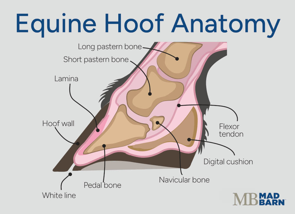 Illustration: Dr. Ana Mesa, PhD
 The epidermis comprises both the outer hoof wall — made up of hardened, keratinized material — and the living cells that lie beneath it. The hoof wall is responsible for bearing the majority of the horse’s weight by transferring force from the ground to the underlying bone. [1][5]
Hoof Laminae
Within the hoof, the dermis and epidermis interlock through an intricate system of lamellae (a.k.a laminae), which are finger-like projections that anchor the hoof wall to the underlying bone. [1]
There are about 550 – 600 primary epidermal lamellae surrounding the coffin bone, each of which branches into hundreds of smaller secondary lamellae. These interwoven layers form a strong, Velcro-like connection that provides stability and support. [1]
Between these two interlocking sets of lamellae lies the basement membrane – a thin membrane that helps to anchor the hoof wall to the coffin bone. [7]
Effects of Laminitis
In cases of laminitis, the attachment system between the epidermal and dermal lamellae begins to break down in response to prolonged inflammation.
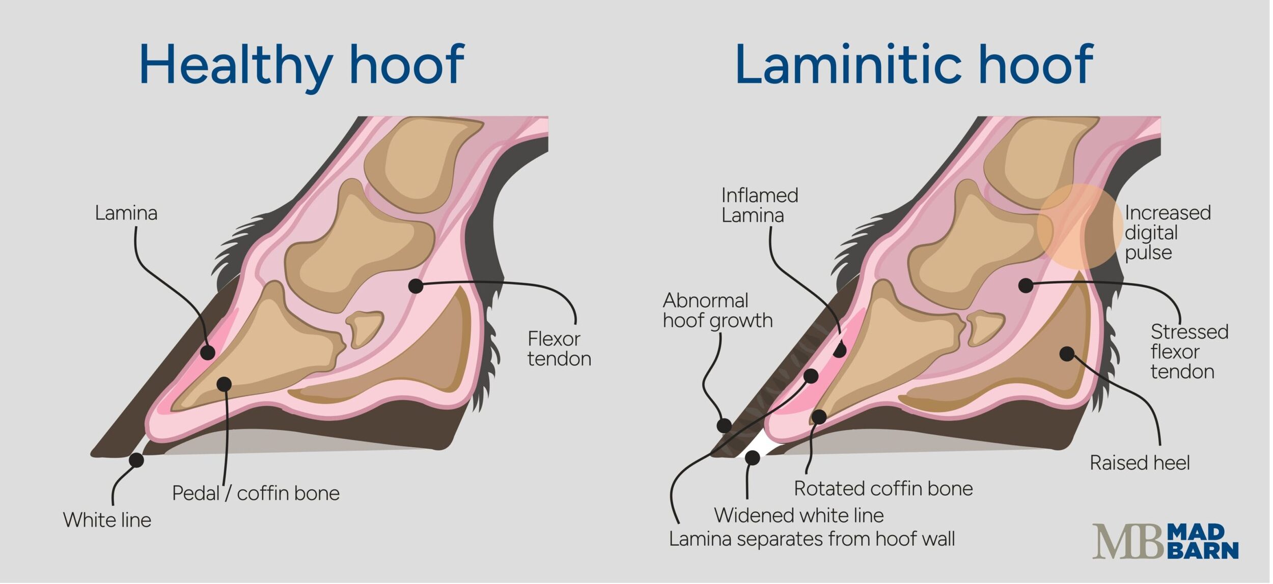 Illustration: Dr. Ana Mesa, PhD
 Inflammation disrupts the cells’ internal structure, impacting their ability to adhere to the basement membrane. Over time, the lamellar epithelial cells become weakened. This leads to stretching and, in severe cases, separation of the epidermal and dermal lamellae. [1]
When this happens, the coffin bone loses its firm connection to the hoof wall, causing instability, pain, and in extreme cases, rotation or sinking of the bone within the hoof. [1]
This disease process describes all forms of laminitis. In cases of supporting limb laminitis, inflammation in the lamellae is caused by prolonged excessive force placed on one limb. [1]
Symptoms
As with all forms of laminitis, the characteristic symptom of SLL in horses is lameness. [5][6]
SLL is challenging to identify because affected horses are usually already lame due to the underlying condition that led to compensatory weight bearing in the first place. [6]
A frequent indicator of supporting‑limb laminitis (SLL) is a sudden, noticeable reduction in lameness in the originally injured limb. This apparent improvement usually occurs because the weight‑bearing limb also becomes painful, prompting the horse to shift more weight to whichever limb currently hurts less.
However, in some cases the original injury or infection in the opposite leg has been resolved for weeks or even months before SLL develops. [8]

“Early recognition, use of anti-inflammatory medications, and supporting the affected foot with soft substrates, alongside careful management of the primary injury, are essential to prevent catastrophic hoof rotation in cases of supporting limb laminitis.”
— Dr. Jennifer Skaggs, DVMEquine Veterinarian
The outward symptoms of laminitis appear after internal inflammation has already occurred. This increases the difficulty of diagnosing it early enough for treatment to be effective. [1]
Symptoms of SLL include: [1][3]
- Shifting the weight from foot to foot
- Increased heart rate
- Increased digital pulse in the affected hoof
- Increased temperature in the affected hoof
- Changes to the coronary band of the affected hoof
What's your top priority with your horse's health?

What's your top priority with your horse's health?
Causes
The causes of SLL have not been completely characterized at this time. Currently, there is evidence that SLL is a complex condition influenced by: [3][7]
- Blood flow restriction
- Cellular stress
- Glucose supply
- Inflammation
- Mechanical forces
Together, these forces contribute to progressive breakdown of the hoof’s support structures. [3][7]
When a healthy horse puts weight on its hoof, blood flow in certain parts of the foot decreases or stops. [3] The deep digital flexor tendon blocks blood flow by pulling on the coffin bone.
As weight is applied, the tendon flattens against bones in the lower leg, which puts pressure on nearby arteries. Other structures, such as ligaments and connective tissue, may also contribute to this effect. [3]
This temporary loss of blood flow during normal movement is a natural function, preventing backflow and regulating circulation. [3] Healthy horses simply shift their weight back to the opposite leg, allowing blood flow to resume.
In cases where a horse has an existing injury, it may favor the painful leg, keeping weight on the uninjured hoof for a prolonged period. This prevents restoration of normal blood flow through the hoof. [6]
Without normal blood flow, changes occur in the lamellae related to lack of oxygen and glucose. This leads to cell stress, weakening of the tissues, and eventually tissue death. The cells located at a greater distance from the blood supply are impacted first, leading to structural damage in the hoof. [3][6]
Tissue samples from horses with SLL reveal cellular responses to inflammation that are similar to inflammatory skin diseases like psoriasis. This suggests the condition involves not only mechanical stress from excess weight bearing but also inflammation and disrupted cell function. [6]
Risk Factors
Horses that experience injuries, lameness, or pain in one or more hooves are at greater risk of developing SLL. [6] This is because an injury in any hoof results in compensatory movement and load bearing. [6]
Possible causes of pain in the hooves include: [6]
- Bone fractures within the hoof capsule (e.g., distal phalanx/coffin bone, navicular bone, distal sesamoid)
- Hoof infections such as abscesses or white‑line disease
- Orthopedic injuries that require casting
- Post‑surgical recovery (e.g., following fetlock arthrodesis)
SLL is a common complication in horses recovering from hoof injuries. [6]
In cases where an existing injury has caused lameness, the development of SLL sometimes goes unnoticed, since it is difficult to differentiate lameness related to the injury from lameness due to compensation. [6]
Horses carrying extra body weight are also at increased risk of developing SLL. [6] SLL is more likely to develop in adult horses than in foals or yearlings. [3]

Diagnosis
Diagnosis of SLL is based on the presence of lameness alongside a preceding injury in the opposite leg. [6] Veterinarians may use imaging (X-ray), lameness exams, and flexion tests to confirm diagnosis. [1]
Diagnosing supporting limb laminitis may be challenging because the lameness it causes can be difficult to distinguish from the original injury. [6] Additionally, while the noticeable symptoms of SLL tend to come on quickly, they usually occur after early changes have taken place in the hoof, making early diagnosis difficult. [1]
Treatment & Prognosis
Treatment of SLL in the chronic phase may include supporting the affected hoof with the use of corrective farriery, which may include special shoes or orthotics. [6][7] It is also necessary to resolve the injury or health condition that led to compensation in the first place.
Treatments used in the acute phase may include: [5][6][7][8]
- Pain relief
- Anti-inflammatory medications
- Cryotherapy (ice boots)
- Digital blood flow therapy
- Stall rest
- Nutrition interventions to support hoof growth
The prognosis for horses with SLL is poor. [6] This is particularly true in cases where the injury in the opposite limb remains unresolved. [6]
Prevention
Preventing SLL after hoof injury is challenging. Research trials of therapeutic shoes, modified stall surfaces, and orthotic devices have yet to show consistent benefit. [6]
Prevention of SLL requires careful monitoring of horses with injured or infected legs or hooves.
Treatments that help horses stay mobile or encourage them to shift their weight between the limbs after hoof or leg injury are recommended. [6][7]
While SLL is difficult to prevent once the horse has already sustained a limb injury, optimal management practices can help avoid situations that pose a high risk of injury. Strategies include:
- Use appropriate footing: Make sure training areas are well maintained and suitable for the type of work your horse is doing
- Warm up and cool down: Allowing your horse time to warm up and cool down before intense exercise is key to building fitness and preventing injury
- Optimize recovery: The rest period between workouts gives your horse’s muscles time to repair and build stronger tissue
- Manage pasture: Routine maintenance and regular hazard checks go a long way to preventing horse injuries
Finally, a well-balanced, forage-first diet is key to equine weight management and overall health. If you need help creating a plan tailored to your horse’s individual requirements, book a free consultation with one of our qualified equine nutritionists.
Frequently Asked Questions
Here are some frequently asked questions about supporting limb laminitis in horses:
Supporting limb laminitis is a serious form of laminitis that develops when a horse overloads one limb after pain, injury, infection, or surgery affects the opposite leg. Prolonged uneven weight bearing can reduce blood flow inside the supporting hoof, stressing the laminae that hold the hoof wall to the coffin bone. Severe cases can progress to founder, coffin bone rotation, or sinking.
Supporting limb laminitis usually occurs after a horse avoids using one painful limb and keeps too much weight on the opposite leg. Normal weight shifting helps restore hoof blood flow, but prolonged loading can restrict circulation, reduce oxygen and glucose delivery, and trigger cellular stress. Over time, inflammation and mechanical strain weaken the hoof’s internal support structures.
Early signs of supporting limb laminitis can include new or worsening lameness in the supporting limb, a stronger digital pulse, increased hoof heat, and frequent weight shifting. Some horses also show an increased heart rate or changes around the coronary band. A sudden improvement in the originally injured limb can also be a warning sign if the opposite limb has become painful.
Supporting limb laminitis is a veterinary emergency because damage inside the hoof can progress quickly once blood flow and laminar attachment are disrupted. Delayed treatment increases the risk of founder, where the coffin bone separates from the hoof wall and may rotate or sink. Horses showing possible signs need prompt veterinary assessment and pain control.
Horses at higher risk of supporting limb laminitis include those recovering from hoof injuries, fractures, infections, orthopedic surgery, or conditions that require casting. Adult horses and overweight horses may also face greater risk. Any painful limb problem that causes prolonged compensation can increase stress on the opposite supporting hoof.
Treatment for supporting limb laminitis focuses on reducing pain, supporting the affected hoof, limiting inflammation, and resolving the original injury that caused uneven weight bearing. Veterinary care may include anti-inflammatory medication, pain relief, cryotherapy, digital blood flow therapy, stall rest, corrective farriery, special shoes, or orthotics. Nutrition support may also help maintain hoof growth during recovery.
Founder can develop from supporting limb laminitis when inflammation severely weakens the laminae that anchor the hoof wall to the coffin bone. As this support system fails, the coffin bone can lose stability and shift within the hoof. Severe founder is painful, difficult to treat, and may be life-limiting in some horses.
Prevention of supporting limb laminitis depends on close monitoring after limb injuries and helping the horse avoid prolonged overload on one foot. Veterinary-guided pain control, supportive footing, appropriate hoof support, and treatment of the original injury may help reduce risk. Owners should watch for hoof heat, stronger digital pulses, shifting weight, or new lameness.
Summary
Supporting limb laminitis (SLL) in horses is caused by prolonged weight-bearing on a single limb. It often develops in horses with an existing injury or lameness in one limb, forcing them to shift excessive weight onto the opposite hoof.
- The characteristic symptom is lameness. Other symptoms include increased digital pulse, hoof temperature changes, shifting weight between feet, and coronary band changes
- Horses with injury, fractures, or infections in a hoof or leg are at risk of developing SLL
- The cause of SLL is prolonged weight-bearing on one limb due to injury or infection in the opposite leg
- Treatment requires supportive shoeing along with pain relief, anti-inflammatories, cryotherapy, and stall rest
- The prognosis for SLL in horses is poor, especially if the original injury persists
- Prevention focuses on balancing weight redistribution during recovery from leg injury or infection, mobility support, and early intervention
References
- Stashak. T. S. and Baxter. G. M., Adams and Stashak’s Lameness in Horses. 7th edition. wiley Blackwell, Hoboken. 2020.
- Elliott. J. and Bailey. S. R., A Review of Cellular and Molecular Mechanisms in Endocrinopathic, Sepsis‐related and Supporting Limb Equine Laminitis. Equine Veterinary Journal. 2023. View Summary
- Orsini. J. A., Supporting Limb Laminitis: The Four Important 'Whys'. Equine Veterinary Journal. 2012. View Summary
- Wylie. C. E. et al., Prevalence of Supporting Limb Laminitis in a UK Equine Practice and Referral Hospital Setting between 2005 and 2013: Implications for Future Epidemiological Studies. Veterinary Record. 2015. View Summary
- Ross. M. W. and Dyson. S. J., Eds., Diagnosis and Management of Lameness in the Horse. 2nd ed. Elsevier/Saunders, St. Louis, Mo. 2011.
- Van Eps. A. et al., Supporting Limb Laminitis. Veterinary Clinics of North America: Equine Practice. 2021. View Summary
- Van Eps. A. W. and Burns. T. A., Are There Shared Mechanisms in the Pathophysiology of Different Clinical Forms of Laminitis and What Are the Implications for Prevention and Treatment?. Veterinary Clinics of North America: Equine Practice. 2019. View Summary
- Hopster. K. and Driessen. B., Pharmacology of the Equine Foot. Veterinary Clinics of North America: Equine Practice. 2021. View Summary