


Radiographs, commonly known as X-rays, are one of the most valuable diagnostic tools in equine veterinary medicine. These images allow veterinarians to examine the internal structures of the horse, primarily bones and joints, without the need for surgery or invasive procedures.
Whether evaluating a persistent lameness, investigating swelling or pain, or conducting a prepurchase exam, radiographs provide critical information that can guide treatment decisions and help monitor healing over time.
Proper patient preparation and positioning are essential to obtaining clear and accurate images. A calm, cooperative horse, a clean imaging site, and a skilled veterinary team all contribute to successfully generating clear radiographic images. Sedation may be necessary to keep the horse still during imaging, which helps ensure high-quality images and safety for the veterinary team.
Read on to explore how radiographs are used in equine medicine, how to prepare a horse for radiographic examination, and what to expect during the process. Whether you’re a horse owner, trainer, or equine professional, understanding the role radiographs play in diagnosis and treatment can help you make informed decisions about your horse’s health and performance.
Radiography for Horses
Radiographs, also called X-rays, are an important diagnostic tool for evaluating the bones and joints of horses. They are most commonly used when working up a lameness case, as they allow the veterinarian to detect conditions such as arthritis or osteochondrosis dissecans.
Radiographs are images produced by directing X-ray photons, a form of radiation, through the horse’s tissues onto a digital plate. Each tissue absorbs a specific amount of X-ray energy, limiting how much makes it through to the other side and onto the plate. [1]
The densest tissue, bone, absorbs nearly all of the energy directed at it, preventing the X-rays from reaching the film or plate. These areas show up as white on the X-ray. In contrast, bodily fluids absorb very little energy, so most of the photon’s energy makes it to the plate. These areas appear black on the final image. [1]
Veterinarians can then interpret the final image to identify any changes that may explain a horse’s lameness.
Clinical Use
Veterinarians use X-rays to evaluate a horse’s bones and joints. In some cases, they may also use radiographs to look for specific types of soft tissue abnormalities.
Common uses for X-rays include: [1]
- Diagnosis of lameness conditions affecting the limbs, neck, or back
- Pre-purchase examinations
- Evaluating for dental conditions, such as equine odontoclastic tooth resorption and hypercementosis (EOTRH)
- Assessing for fluid in the nasal sinuses caused by sinus infections
- Looking for sand in the abdomen when working up chronic colic cases
What's your top priority with your horse's health?
Enter your email to receive your store credit
Equipment
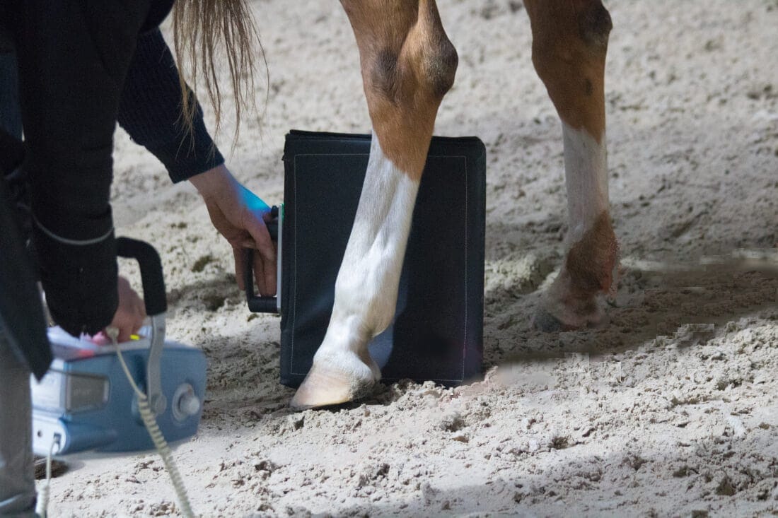
There are two basic parts to a veterinary X-ray system: the X-ray generator and the cassette. [1]
X-ray generators produce the photons recorded by the cassette. Many equine practitioners use mobile X-ray generators, which allow them to take radiographs on the farm. These units are portable and only require a power supply to operate. However, they are less powerful than the X-ray machines available in-clinic. This makes them more susceptible to image blurring. It also makes taking suitable images of the shoulder, pelvis, and other structures difficult, as the machine is not powerful enough to send photons through the thicker tissues. [1]
The cassette holds the digital plate that records the X-ray image. For portable X-ray systems, an assistant holds the cassette behind the area being X-rayed, making sure it is aligned with the beam of X-rays coming from the generator. Movement of the cassette when shooting the X-ray can lead to a blurred image. [1] The veterinarian can also tape metal icons to the cassette to produce a label on the final image. Typically these are a metal “L” or “R”, to indicate whether the image is of the left or right leg. [1]
When taking images of the hooves, foot-positioning aids are necessary to ensure that the cassette is in the correct location. Some X-ray systems require the cassette to be placed under the horse’s foot to generate hoof images. In this case, the veterinarian slides the cassette into a protective box that still allows photons to reach the plate. [1]
Safety
Exposure to X-ray radiation can cause significant health issues in people, particularly when they are exposed to radiation frequently or at high levels during a single exposure. Given this, taking proper safety precautions is vital for the veterinary team, many of whom take X-rays daily.
Safety equipment required for anyone in the vicinity of an active X-ray machine includes: [1]
- Lead-lined gowns, gloves, and thyroid guards
- Cassette holders to increase the distance between the assistant and the cassette
- A radiation dosimeter, which measures the amount of radiation exposure a person has had
People who work with X-rays are also required to take specialized nuclear safety training and register with the government for tracking of their radiation exposure over time.
X-Ray Procedure
The process of taking a radiograph is relatively simple; however, proper preparation is key to ensuring a clear diagnostic image.
The most common reasons repeated X-rays are required for horses are: [1][2]
- Movement blur: From the horse or the X-ray machine moving during shooting
- Poor positioning: Due to misalignment of the X-ray machine or an uncooperative horse
- Artefacts: Materials that the X-ray was not meant to capture but interfere with photon absorption
In addition, young or pregnant people should not be exposed to X-rays.
To avoid movement blur, veterinarians typically sedate the horse to limit their movement. The type of sedative used depends on the individual horse and the number of X-rays that need to be taken. Using cassette holders, blocks, or other objects to rest the cassette and generator on can also help minimize movement blur.
Proper positioning often requires the horse to stand in a particular manner, such as up on a foot block or with their limb held so they are non-weight-bearing. [1] Sedation can help improve positioning by making the horse more cooperative. It also gives the veterinarian time to align the X-ray generator and cassette properly, without having to worry about the horse moving out of alignment.
Many artefacts on equine X-rays are due to debris, dirt, clumps of wet hair, or other foreign materials on the horse’s skin. [1] Grooming the horse to remove debris can increase the quality of the X-ray images. [1]
For radiographs including the hoof, veterinarians typically pack the hoof using plasticine or a similar material. This material fills any cracks or gaps in the horse’s hoof, preventing air pockets in these areas from casting shadows over the bones that may be misinterpreted. [1]
Once the horse is ready for the procedure, the veterinarian lines up the X-ray generator and the cassette (held by their assistant) to image the area in question. After activating the X-ray generator, the machine beeps and the image is ready to be examined. For digital X-rays, the cassette transfers the information to a computer, where the radiograph can be viewed immediately. For analog X-rays, the cassette film must be developed before viewing, but this method is less common with the widespread use of digital equipment. [1]
For each area examined, the veterinarian takes at least two images, 90 degrees apart. This is called an orthogonal view, and allows the veterinarian to cross-reference any abnormalities seen in one image with the same image from a different angle. This is particularly important for conditions like fractures, where an artefact may cause the appearance of a fracture in one image, but is not visible when the area is viewed from another angle.
Interpreting Results
Interpretation of radiographs can be challenging, as they produce a two-dimensional image of a three-dimensional object. Bony structures, soft tissues, and other features overlap in the final image, making them difficult to accurately distinguish from each other.
To evaluate the radiograph, the veterinarian goes through the image systematically to ensure that all bones and joints are examined. They may compare the images to radiological atlases, textbooks that contain images of normal and abnormal bones and joints. [1] Adjusting the contrast of the image can help identify subtle changes that may be missed otherwise. [1]

“Radiographs are one of our most powerful tools for diagnosing lameness and bone injuries. As a mainstay of equine medicine, these images typically give us clear answers that guide effective treatment decisions in most cases.”
— Dr. Jennifer Skaggs, DVMEquine Veterinarian
Your veterinarian may recommend sending the images to a diagnostic imaging specialist. These veterinarians receive additional training in interpreting radiographs. If your veterinarian has a digital system, sending X-rays for specialist interpretation is simple using online digital imaging platforms.
It’s important to note that radiographs are typically only suitable for evaluating bony structures. Soft tissues such as ligaments, tendons, and cartilage may appear on an X-ray, but are not shown in sufficient detail to allow for diagnosis.
The main findings on radiographs are either increased bone or bone loss. [1] Both processes indicate bone disease that may cause lameness in horses. Veterinarians can also identify fractures using X-ray images.
Increased Bone
Most cases of increased bone are areas of new bone formation, such as at the attachment sites of ligaments or around a joint. This is an indicator of long-term inflammation or damage to the bone, such as from degenerative arthritic changes. [1]
These areas typically have a rough, irregular appearance on X-rays. A smooth area of new bone proliferation may indicate a previous injury that is no longer causing lameness or pain. [1]
Bone can also become sclerotic (more dense). Sclerotic bone is more white than normal bone and indicates that the bone is under strain. [1] For example, the front portion of the cannon bone is denser than the back, as it comes under more strain during normal movement. Abnormal sclerosis can indicate additional wear-and-tear or stress on a particular area.
Decreased Bone
Bone loss occurs either due to resorption or lysis (tissue destruction). Resorption is the body removing bone as part of its ongoing bone maintenance processes. In most cases, resorption occurs when there is pressure on the bone surface. For example, when there is a keratoma, a mass within the hoof capsule, the mass can put pressure on the coffin bone and cause resorption. Resorption typically has smooth edges in X-ray imaging and occurs slowly over time. [1]
Lysis suggests there is active destruction of bone by some other process, such as a bacterial infection. It typically has rough, irregular edges in imaging and occurs more quickly than resorption. [1]

Fractures
Fractures are a crack or gap in the bone that is visible in X-ray images. They appear as a dark line within the typically white or gray bone. There are many different types of fractures, and they are classified based on the type of fracture, severity, and the size of the gap between the bone fragments. [3]
During the initial stages following a fracture, it may be very difficult to see on a radiograph. For the fracture to appear, the bone fragments must separate from each other slightly. Non-displaced fractures, where the bones are still touching each other in a normal configuration, may not be visible on X-rays. It can take 5 – 10 days for the fracture to become visible following this type of injury. [3]
Preparing for Radiographs
Having radiographs taken requires minimal preparation. Before your veterinarian arrives, make sure your horse is clean and has their hooves picked out. Position your horse in a quiet, safe area with a power supply available. Barn aisles are often ideal locations for taking X-rays.
If your veterinarian has asked you to give an oral sedative, such as detomidine gel, give the medication according to their timing and dosage instructions.
If your horse is shod, your veterinarian may ask you to have the horse’s shoes removed prior to taking X-rays. Metal shoes can interfere with the X-ray beam when taking images of the hoof. They can also block key structures that need to be examined. Coordinate with your farrier to have your horse’s shoes taken off in time for the appointment.
Complications
X-rays are very safe for the horse, as they receive only a small amount of radiation during the procedure. Rarely, complications may arise from horses reacting to sedation used to keep them still during imaging.
Most adverse effects are mild and short-lived, and with routine monitoring and supportive care, horses recover uneventfully.
Frequently Asked Questions
Here are some frequently asked questions about radiographs for horses:
Radiographs are used to view the horse's bones and joints, helping diagnose fractures, arthritis, laminitis, bone cysts, OCD, hoof problems, dental issues, and more.
Horses do not always need to be sedated for X-rays. Many horses tolerate radiographs well without sedation. However, nervous or fidgety horses may need mild sedation to stay still so the veterinarian can obtain clear images.
For hoof radiographs, shoes are often removed to avoid image distortion. Your veterinarian will let you know if shoe removal is necessary. For radiographs elsewhere on the body, the shoes do not need to be removed prior to the procedure.
The radiation dose emitted during a single X-ray imaging session is very low and safe for your horse. The veterinary team will take precautions to limit their own exposure, such as wearing lead aprons and stepping out of the beam. Most veterinary teams ask owners to step out of the area to minimize their exposure to radiation. It is important to communicate with your veterinarian if you or someone at the appointment is pregnant, as exposure to radiation can be harmful to a developing fetus.
Summary
Radiographs, or X-rays, are a valuable diagnostic tool for assessing a horse's bones and joints.
- X-rays can help diagnose bony changes, including new bone formation or bone loss
- Most X-rays are used for diagnosis of lameness conditions in the hooves and limbs
- Some horses may require sedation for the procedure, depending on their personality and the type of images taken
- The procedure is safe for the horse due to the low dose of radiation
References
- Weaver. M. and Barakzai. S. Handbook of Equine Radiography. Saunders Elsevier, Edinburgh. 2010.
- O'Brien. T. O'Brien’s Radiology for the Ambulatory Equine Practitioner. Teton NewMedia, New York. 2005.
- Janet A. Butler et al. Clinical Radiology of the Horse. Wiley-Blackwell, Chichester, West Sussex, UK. 2016.


