


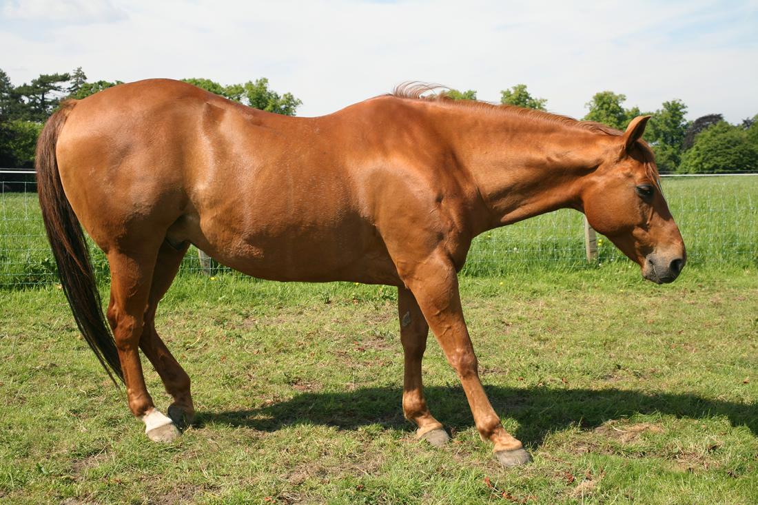
Founder is the common name for laminitis, a condition defined as inflammation of the laminae within the hoof. It can cause lameness in horses, ponies and donkeys and involves damage to the laminar connection between the hoof wall and the coffin bone.
This may lead to rotation and/or sinking of the coffin bone, which causes severe pain and can permanently damage the hoof structure. Horses with the condition may show various signs of distress and discomfort and typically cannot move about comfortably.
The term founder is often used in the context of a horse that has chronic (long-term) or repeated laminitis episodes, but horses can have a single episode of founder or laminitis.
There are multiple risk factors for the development of founder, but over 90% of cases are caused by high insulin (hyperinsulinemia), Equine Metabolic Syndrome (EMS) and Cushing’s disease (PPID). Many other risk factors are involved and will be discussed in further detail below.
Prevention and treatment of founder involve addressing the underlying causes of the condition and require a lot of dedication from the horse owner. Founder is not always treatable if the internal structures of the foot have sustained significant damage but the vast majority can be saved with elimination of the cause and correct hoof care.
Proper feeding, hoof care, and management are critical for supporting hoof health. If your horse is recovering from or at risk of founder, our nutritionists can help you develop a balanced feeding plan to address laminitis risk and optimize hoof health.
What is Founder?
A debilitating and painful condition, founder affects the laminae and the coffin (pedal) bone in the equine foot.
The laminae are the two layers of finger-like protrusions that interlock or interdigitate with each other like Velcro to form the structure that holds the hoof wall onto the internal structures. The laminae also help support the coffin bone.
The coffin (pedal or P3) bone is the bone that rests in the cradle of the hoof, and it plays a key role in hoof form and function. It is the first bone in the boney column of the leg to absorb the shock of the footfall, and its proper function is critical to limb circulation. [1]
 Illustration: Dr. Ana Mesa, PhD
 To confirm a diagnosis of founder, your veterinarian will conduct clinical and radiographic examination of the hooves and the entire horse. [1] In the physical exam, your veterinarian will look for signs of current and chronic (ongoing) laminitis while also investigating hoof structure and causes for the episode.
Radiographs show the degree of coffin bone rotation, coffin bone sinking (also known as distal descent), sole and wall thickness, toe length and, sometimes, concurrent abscesses. This information can help your veterinarian, farrier and nutritionist work together to relieve your horse’s pain with corrective foot care, feed changes, housing changes and medications.
Laminitis vs. Founder
The terms “laminitis” and “founder” are often used interchangeably by horse owners. However, a distinction can be made between the acute phase of laminitis versus ongoing chronic issues.
Acute laminitis or a “bout of laminitis” typically refers to the sudden onset of pain related to weakening of the laminae.
The term founder is often used colloquially to refer to the ongoing condition when it involves rotation/sinking of the coffin bone. [2]
Not all horses with acute laminitis will experience coffin bone rotation. However, many horses with founder will have previously had one or more attacks of laminitis. Once a horse has had one episode, it is usually at risk of reoccurrence.
Laminae
Each horse hoof contains approximately 600 laminae forming two interlocking layers that provide structural support to the hoof. The laminae connect the hoof wall to the coffin bone within the hoof.
When the laminae become weakened, the following can result:
- Separation of the sole from the hoof wall observed at the white line of the hoof; the area between the wall and sole of the hoof
- Flat hoof sole
- Divergent hoof rings
- Acute Laminitis
- Founder/Chronic Laminitis
What's your top priority with your horse's health?
Enter your email to receive your store credit
Acute Laminitis
During acute laminitis episodes of non-metabolic causes, several factors contribute to damage to the laminae. Toxins can activate matrix metalloproteinase enzymes. The tissues are invaded by white blood cells in an inflammatory response. Blood supply is compromised either as an initial event or later on secondary to tissue swelling and clot formation.
In metabolic / endocrinopathic laminitis, the inflammatory features of white cell infiltration and enzyme activation are not present but there are elevated levels of the potent vasoconstrictor endothelin-1 suggesting blood flow is compromised. [15]
A laminitis attack can lead to the death of cells in the laminae as well as cell proliferation. If there is enough damage, the laminae lose their strength, weakening this important structural component of the hoof.
In a hoof affected by founder, the laminae can become so damaged that they are unable to sufficiently anchor the coffin bone. The position of the coffin bone may drop (sink) under the weight of the horse and due to the upward pull of the flexor tendon attached to the base of the bone. [1]
This is known as coffin bone rotation. The degree of rotation is measured by taking radiographs of the hoof.
In advanced cases of founder, the coffin bone can protrude through the sole of the hoof. [1] At this stage, euthanasia is often considered because of the degree of nursing and prolonged period required for recovery but horses can recover from penetration.
Effects of Founder on the Foot
When rotation of the angle of the coffin bone occurs relative to the short pastern bone located above it, intense pressure is exerted on the sole of the foot potentially causing it to penetrate the sole.
A depression may be present at the front of the coronet where the joint space between the coffin and pastern bone becomes wider than normal. [4]
Prevalence and Prognosis
Half of all US horse operations surveyed in 1998 reported having at least one horse with lameness and 13% reported having a horse with laminitis.
The survey also found that 4.7% of horses with laminitis died or had to be euthanized due to the condition. [3]
In this owner-reported survey, over 50% of reported laminitis cases were thought to be due to grazing on lush pasture and grain overload.
Up to 74% of horses with laminitis recovered and were able to be used for their intended purposes.
A British study of equine veterinary clinics found that active laminitis cases represented one out of every 200 visits to veterinary practitioners. [20]
Common Causes of Founder
Founder is a complex condition that can be precipitated by both metabolic and environmental factors acting on their own or in combination with each other. [4]
Some of the most common causes of founder include the following.
Dietary Factors
The overconsumption of grain products as in the “horse broke into the feed room” scenario, and experimental overloads of chicory fructan can cause laminitis and founder.
Grains are high in hydrolyzable carbohydrates (HC), including sugar and starch. High intake of starch and sugar can overwhelm the small intestine and lead to excess carbohydrates reaching the hindgut, often called “starch overload”. A similar scenario can be created experimentally with unnaturally large doses of pure chicory root fructan given by stomach tube.
This leads to higher levels of lactic acid production in the cecum and colon. If the pH drops below 6, a condition known as hindgut acidosis develops.
An excessive amount of lactic acid in the hindgut can disrupt the microbial population and cause the death of fibre-fermenting hindgut microbes. As acid accumulates in the intestine, it also causes damage to the gastrointestinal lining. [21][22][23]
However, this is an extreme scenario requiring large overloads and is accompanied by fever, colic and diarrhea. Much smaller amounts of hydrolyzable carbohydrates can cause laminitis by increasing insulin in horses with EMS or PPID.
Absorption of Dietary Toxins
Horses with a compromised gastrointestinal lining absorb toxins into their bloodstream. A common source of toxins is bacterial exotoxins. [15]
Upon entering the bloodstream, these toxins lead to activation of matrix metalloproteinases (MMPs) that damage the hoof laminae.
Research investigating metabolism-related causes of laminar damage suggests that a sugar metabolite called methylglyoxal (MG) may be responsible for damage in the feet of horses. However, a study comparing horses with hyperinsulinemia to normal horses found no difference in levels of MG. [25]
MG produced during the digestive process in horses could be absorbed into the bloodstream and alter the structure of the hoof laminae although there is no actual evidence to suggest this occurs and MG is a very toxic compound which would surely produce other signs. [5]

Metabolic Disorders
Hormonal imbalances including Equine Metabolic Syndrome (EMS) and Cushing’s Disease (Pituitary Pars Intermedia Dysfunction) are associated with an increased risk of laminitis.
In a study of horses that presented at a Finnish veterinary hospital due to laminitis, 89% had evidence of underlying endocrine disorders. One-third were diagnosed with PPID and the remainder had high insulin levels indicative of insulin resistance but without signs of PPID. [6]
Insulin resistance is particularly high among overweight/obese horses, of which 95% have high levels of insulin but without signs of PPID. [6] However, other studies show lower incidence of insulin resistance among overweight/obese horses. It is well recognized that insulin resistant horses may be of normal weight with only regional adiposity at the crest, withers or tailbase. [7][27]
Overweight/obese horses are at higher risk of developing laminitis because a higher percentage of obese horses have metabolic syndrome which also predisposes to easy weight gain.
EMS involves increased insulin secretion from the pancreas and lower movement of glucose into tissues. Elevated levels of insulin in the blood can induce laminitis in horses and ponies. [8][9]
Cushing’s Disease affects the pituitary gland of horses typically over the age of 15. Clinical signs of the disease include an excessively long coat that fails to shed out in the summer, depression, muscle wasting, increased water consumption and urination.
In early cases, unexplained suspensory ligament breakdown and seasonal issues with increased drinking and urination, as well as unexplained first time in life laminitis occur because of an exaggerated seasonal rise in ACTH without the classical coat changes.
Horses with Cushing’s are at high risk of developing laminitis. This is related to higher levels of cortisol and insulin. [10]
Working with a qualified nutritionist to formulate rations for these horses is a life-saving procedure. Simply restricting feed can worsen metabolic conditions, so it is important to design a balanced nutrition plan for your horse or pony.
Testing for Cushing’s Disease (PPID) and EMS is readily available through your veterinarian by taking blood samples and sending them to a laboratory. Blood tests usually look at endogenous ACTH, Glucose and Insulin levels, but more tests may be recommended if other differential diagnoses are being considered.
Acute Illnesses
Health conditions including colic, diarrhea, grain overload, and inflammation in the small intestine (enteritis) affect the horse’s gastrointestinal system.
These conditions may also predispose a horse to develop laminitis and subsequent founder due to the effects of toxins on the laminae.
Endotoxemia occurs when endotoxins (toxins that are produced inside bacterial cells) pass through compromised intestinal tissue and enter the bloodstream. Once in the peripheral blood supply, these toxins can trigger a systemic inflammatory response. [11][12] Bacterial exotoxins from intestinal bacteria can trigger breakdown of the basement membrane of the laminae. [26]
Mares that retain their placenta after giving birth develop bacterial infection in their uterus (metritis) and are at risk for endotoxemia.
If a mare does not pass her full placenta within 3 hours, it is considered abnormal. If she does not pass her placenta within 12 hours, it is considered a medical emergency, and you should contact your veterinarian immediately.
Conditions such as pneumonia and diarrhea in foals and adults due to bacterial infections (i.e. Salmonella, Clostridia, and Potomac Horse Fever) can also cause the release of endotoxins into the bloodstream. Other infections that can result in laminitis include “bastard” (systemic) Strangles and Lyme Disease.
Overweight Body Condition
All equines can be affected by laminitis and founder regardless of their weight.
However, horses and ponies that are overweight are more at risk of pedal bone rotation and damage to their hooves because of the extra weight their feet must support.
It is important to regularly body condition score your horse and adjust their diet if they are carrying excess body fat.
Black Walnut Shavings
The use of black walnut (Juglans nigra) shavings as bedding is a known cause of laminitis After the horse is exposed to black walnut shavings via oral ingestion or skin contact, laminitis may develop within 24 to 48 hours. [13]
Oral ingestion of black walnut extract is a commonly used research technique to induce laminitis in equids. Ingestion of this substance alters blood flow to the hoof and has systemic effects on respiratory function and heart rate. [19]
Impaired Blood Supply in the Foot
Compromised blood supply to the hoof and the laminae within is believed to be a component of many different types of laminitis, including:
- Laminitis due to systemic inflammatory response
- Endocrinopathic laminitis with elevated endothelin-1
- Supporting leg laminitis when a horse is protecting a painful limb by overweighting the opposite leg
Signs and Symptoms of Founder
Laminitis and founder typically affect the front feet of horses but can occur in any of their four feet. [4]
Some of the most common clinical indications include:
- Sudden onset of bilateral lameness
- Resistance to walking or moving
- Feeling an abnormally strong pulse over the paired arteries on the back of the fetlock (the ankle) and heat in the foot
- Shifting weight back and forth between legs
- Reluctance to pick up the feet
- Standing with the legs camped out in front of the body or with all four legs under the body
- Lying down more frequently
Some horses develop chronic laminitis after an episode of acute laminitis. Even with chronic abnormal hoof structure, some horses may not show outwards signs of pain or lameness.
Signs to look out for that can indicate chronic laminitis and impaired hoof integrity include: [14]
- Concavity on the dorsal side of the hoof
- Divergent growth rings (rings of growth on the hoof that are very tight at the toe and wide at the heel)
- Widened white line
- Foot abscesses that develop a few weeks after the initial acute laminitis episode
 Sawhorse Stance Illustration: Dr. Ana Mesa, PhD
 How is Founder Diagnosed?
Owners of horses suspected of having founder should consult with a veterinarian to obtain a precise diagnosis. Horse owners can assist their veterinarian in making a founder diagnosis by providing complete information on their horse’s medical and hoof health history.
When determining if a horse has foundered, a veterinarian will typically complete a thorough physical examination that includes assessing the behaviour, weight, height, temperament, body condition score, body temperature, and vital signs of the horse.
Your veterinarian’s findings on their full physical examination help to determine what other tests they need to determine the best approach for treating your horse or pony.
Lameness Exam
Horses being evaluated for founder typically undergo a lameness examination that includes checking their range of motion, their stance at rest and palpation of their digital pulses (pulses of the vessels on the back of the fetlock).
Hoof testing is also used to determine where the source of pain is in the foot.
However, hoof testers often deceptively show no pain response (a false negative). This can occur when the leg bearing extra weight is experiencing greater pain. This is distracting to the horse and causes them not to react to the hoof tester.
Nerve Block
Nerve blocks may be done if there is any question the pain is coming from the feet or to facilitate procedures like pulling off the shoes, trimming and getting radiographs.
Blood Testing
The most important laboratory testing is to rule out endocrine disease, namely EMS or PPID. ACTH testing for PPID can be done immediately unless pain is very severe. [28]
Insulin testing should be delayed a few days since it is elevated by acute pain.
Stablelab® is a stall-side diagnostic tool that measures Serum Amyloid A (SAA) to test for acute inflammation in the body. Laminitis caused by grass founder or grain overload will not cause an increase in SAA levels. However, if the horse is foundering from another cause, such as infection, SAA will rise. [24]
SAA shows a quick response up and down, so can be useful for the early detection of problems and to see if treatment is working. The Stablelab® test is often repeated every 24 hours. [24]
Radiographs
In horses affected by founder, x-rays are critical for assessing the extent of the damage in the hoof including the degree of hoof rotation that has occurred.
These images are also invaluable to farriers in helping them to choose the most appropriate therapeutic trimming and shoeing strategies to assist the recovery of the horse.
Changes including bone remodelling and rotation of the coffin bone often continue to occur for several months in the hooves of horses affected by founder. X-rays should be taken on a periodic basis to determine if adjustments to the treatment plan are required.
A contrast study, known as a venogram, can be conducted to assess the degree of damage. The veterinarian injects a liquid visible in a radiograph (Omnipaque) into the vein above the hoof.
With a tourniquet placed above the injection site, the veterinarian takes radiographs to assess the blood supply to the laminitic foot. Poor blood supply substantially reduces the prognosis.
A venogram can also be used after farrier care to see if there is any improvement in blood supply.
Treatment of Founder
The sooner that treatment is started, the better the chance of recovery for an affected horse. Appropriate treatment for the condition requires addressing the underlying cause(s).
Emergency Care Practices
In horses that have developed acute founder due to endocrine causes, it is advised to stop feeding all grain products and remove the horse from pasture. [16] Hay should be soaked before feeding until it can be analyzed for hydrolyzable carbohydrate levels.
A foundered horse should have a soft place to stand or lie down to relieve the pressure on the weakened hoof laminae.
Depending on the severity, some horses will need stall rest, whereas others may benefit from being able to move freely to promote blood circulation within the foot.
Learn more in our guide on Emergency Protocols for Acute Laminitis in Horses.
Medication
Your veterinarian will typically administer non-steroidal anti-inflammatory drugs (NSAIDs) to relieve pain and inflammation in horses that have foundered. Devil’s Claw (Harpagophytum procumbens) is an effective herbal alternative.
When additional pain medication is needed, your veterinarians may sometimes prescribe Tylenol and/or Gabapentin. If your horse’s pain is not controlled by one medication prescribed it is important to communicate with your veterinarian. Tylenol has not been well studied in horses and neither has Gabapentin.
Another option is Tramadol, which is free of NSAID side effects and proven effective in laminitis. [29]
Medication to address underlying health conditions, such as PPID and EMS, may also be given and are critical to stopping the process of laminitis. Administering these medications according to your veterinarian’s directions is important for preventing future laminitis episodes and stopping the pain in the current one. Many of these treatments are life-long commitments.
Cold Water Therapy
To reduce inflammation in the foot, a veterinarian may suggest immersing the horse’s foot in ice water for 72 hours to maintain a consistent temperature under 40 degrees Fahrenheit. This is appropriate for grain overload and cases involving systemic inflammation.
In cases of experimentally induced laminitis, keeping the hoof in an ice bath for 48 hours prevented acute laminitis. [17]
Cold water therapy for endocrinopathic laminitis after pain has appeared is controversial because circulation is already impaired.
Cold water therapy reduces blood flow to the area which lowers the amount of toxins and triggering factors that can reach the hoof.
It also inhibits inflammatory pathways in the lamellae which helps maintain a strong connection between hoof wall and coffin bone. [18]
Corrective Trimming and Shoeing
Horses with founder should be made comfortable to bear weight on the rear portion of their affected hooves. This involves a realigning trim with short toe and heels moved back as far as the foot will allow.
Some horses may benefit from being shod with heart bar shoes including therapeutic components such as pads and wedges. Barefoot trimming with boots and pads is usually preferred because the feet often need attention more frequently.
A priority when trimming the hooves of a foundered horse is to remove any flares on the hoof wall to reduce tension on the white line. The walls can be beveled to completely relieve the laminae.
Surgery
Horses with founder may benefit from undergoing surgery on their deep digital flexor tendon (DDFT) to relieve tension on the structure and help prevent rotation of the coffin bone.
However the procedure is controversial and should probably be reserved for cases with severe contraction of the musculotendinous unit.
Even horses which have penetrated the sole or sloughed their hooves can recover but it requires a large investment of time and nursing.
Long-Term Feeding & Management
Horses recovering from endocrinopathic founder require long-term changes in their feeding and management to support healing of damaged tissues and prevent recurrence.
It is recommended to obtain a hay analysis and work with a nutritionist to formulate an appropriate feeding plan.
Follow these tips below to ensure your horse’s diet is appropriate for their needs:
- Feed a forage-based diet and choose mature hay with low levels of hydrolyzable carbohydrates.
- Soak your horse’s hay to remove excess sugars, if needed.
- Eliminate grains or high-energy concentrates in your horse’s diet
- If your horse needs to lose weight, replace some of their hay with low-calorie straw or chaff. However, straw is more prone to moulding than hay and has lower protein and mineral content.
- Use a slow-feeder to provide 1.5% of your horse’s body weight if overweight or 2% of ideal body weight, whichever is larger.
- Turn your horse out on a dry lot or with a grazing muzzle to prevent over-consumption of grass. Some horses will not tolerate grass access, especially in high risk times when the grass is actively growing, such as spring and fall.
- Feed a concentrated vitamin and mineral supplement to ensure your horse gets all of the nutrients required for hoof and metabolic health.
Mad Barn’s AminoTrace+ is a low-NSC vitamin and mineral supplement targeted to the needs of metabolic horses, including those recovering from founder. AminoTrace+ is enriched with higher levels of organic trace minerals and antioxidant nutrients in a low-calorie pellet that is suitable for horses with insulin resistance.

Key Founder Prevention Tips
Proper equine management practices aid in the prevention of founder. Consider the following strategies:
- Regular farrier care: Keeping the hooves in good condition helps prevent stressors that can negatively affect the structure of the laminae.
- Veterinary wellness checks: Horses should be checked and treated for health conditions such as EMS and PPID that may affect their hoof health.
- Dietary monitoring: Horses should be carefully introduced to grass pastures to prevent weight gain that can cause excess weight-bearing on the hooves. Horses with metabolic concerns should not be fed grain or starchy feeds, while others may be safely fed a limited amount of these food sources. Horses with metabolic disease typically require limited or no pasture access.
- Body condition scoring: Owners should get comfortable with regularly monitoring their horse’s body condition score. If your horse starts gaining excess body condition, adjust their feeding program to reduce energy intake.
For help with meeting your horse’s dietary needs, submit their diet online for a free review by our equine nutritionists.
Frequently Asked Questions
Here are some frequently asked questions about founder in horses:
Founder in horses, also known as laminitis, is a painful condition where the laminae within the hoof become inflamed, leading to potential damage to the connection between the hoof wall and the coffin bone. This can result in lameness, severe pain, and in some cases, permanent damage to the hoof structure. [1]
Symptoms of founder in horses include sudden lameness, reluctance to move or pick up their feet, and an abnormally strong pulse or heat in the affected foot. Horses may also shift weight between legs, stand in unusual postures, or lie down more frequently due to pain. [14]
Treatment for founder in horses focuses on relieving pain, reducing inflammation, and addressing the underlying causes. This may include anti-inflammatory medications, cold water therapy, and dietary adjustments such as eliminating grain and feeding low-sugar hay, alongside long-term management to prevent recurrence. [5][17][29]
There is no guaranteed cure for founder in horses, but mild cases may recover with early treatment and proper care. Recovery is often lengthy and uncertain, and severe cases may require euthanasia if pain cannot be adequately managed. [1]
Summary
Founder is painful inflammation of the hoof laminae that can lead to coffin bone rotation/sinking, severe lameness, and permanent hoof damage.
- Over 90% of cases are caused by hyperinsulinemia from EMS or PPID, though diet, toxins, illness, mechanical overload, and obesity can also contribute.
- Typical signs of founder include sudden bilateral lameness, heat and strong digital pulses, reluctance to move, and a “sawhorse” stance, with chronic cases showing divergent rings and a widened white line.
- Vets confirm founder with clinical exams and radiographs to assess rotation/sinking and guide corrective care, often alongside endocrine testing for EMS/PPID.
- Feed a low-NSC, forage-first diet (soaked hay as needed), manage weight and pasture access, supplement balanced vitamins/minerals, and maintain regular farriery and veterinary monitoring to reduce founder recurrence.
References
- Young, A. Laminitis. UC Davis. 2020.
- Laminitis: Prevention & Treatment. AAEP. 2021.
- Lameness and laminitis in US Horses. USDA. 2000.
- Belknap, J.K. and Geor, R.J. Equine Laminitis. Wiley & Sons. 2016.
- Vercelli, C. et al. Effect of sugar metabolite methylglyoxal on equine lamellar explants: An ex vivo model of laminitis. PLoS One. 2021.View Summary
- Karikoski, N.P. et al. The prevalence of endocrinopathic laminitis among horses presented for laminitis at a first-opinion/referral equine hospital. Dom Anim Endocrin. 2011.View Summary
- Geor, R.J. Metabolic Predispositions to Laminitis in Horses and Ponies: Obesity, Insulin Resistance and Metabolic Syndromes. J Equine Vet Sci. 2008.
- de Laat, M.A. et al. Equine laminitis: induced by 48 h hyperinsulinaemia in Standardbred horses. Equine Vet J. 2010. View Summary
- Asplin, K.E. et al. Induction of laminitis by prolonged hyperinsulinaemia in clinically normal ponies. Vet J. 2007. View Summary
- Donaldson, M.T. et al. Evaluation of suspected pituitary pars intermedia dysfunction in horses with laminitis. J Am Vet Med Assoc. 2004. View Summary
- Tadros, E.M. et al. Effects of a “two-hit” model of organ damage on the systemic inflammatory response and development of laminitis in horses. Vet Immunol Immunopath. 2012.View Summary
- Visser, M.B. and Pollitt, C.C. Lamellar leukocyte infiltration and involvement of IL-6 during oligofructose-induced equine laminitis development. Vet Immunol Immunopath. 2011.
- Black Walnut and Butternut Poisoning of Horses. OMAFRA. 2016.
- Onishi, J.C. et al. Chronic laminitis is associated with potential bacterial pathogens in the laminae. Vet Microbiol. 2012. View Summary
- Gauff, F. et al. Hyperinsulinaemia increases vascular resistance and endothelin-1 expression in the equine digit. Equine Vet J. 2013. View Summary
- Watts, K. and Pollitt, C. Equine Laminitis: Managing Pasture to Reduce the Risk. Australian Government. 2010.
- van Eps, A.W. and Pollitt, C.C. Equine laminitis: cryotherapy reduces the severity of the acute lesion. Equine Vet J. 2010.View Summary
- van Eps, A.W. et al. Digital hypothermia inhibits early lamellar inflammatory signalling in the oligofructose laminitis model. Equine Vet J. 2011. View Summary
- Adair III, H.S. et al. Laminar microvascular flow, measured by means of laser Doppler flowmetry, during the prodromal stages of black walnut-induced laminitis in horses. Am J Vet Res. 2000.View Summary
- Wylie, S.N. et al. A cohort study of equine laminitis in Great Britain 2009-2011: estimation of disease frequency and description of clinical signs in 577 cases. Equine Vet J. 2013. View Summary
- Rowe, J.B. et al. Controlling acidosis in the equine hindgut. Advances in Animal Nutrition in Australia. 1995.
- Destrez, A. et al. Changes of the hindgut microbiota due to high-starch diet can be associated with behavioral stress response in horses. Physiol Behav. 2015. View Summary
- Davies, J. et al. Development of electrochemical DNA biosensor for equine hindgut acidosis detection. Sensors. 2021. View Summary
- Stablelab. Zoetis. Accessed 03/22/2023.
- Ragno, V.M. et al. Morphometric, metabolic, and inflammatory markers across a cohort of client-owned horses and ponies on the insulin dysregulation spectrum. J Equine Vet Sci. 2021. View Summary
- Mungall, B.A. et al. In vitro evidence for a bacterial pathogenesis of equine laminitis. Vet Microbiol. 2001. View Summary
- Stefaniuk-Szmukier, M. et al. Equine Metabolic Syndrome: A Complex Disease Influenced by Multifactorial Genetic Factors. Genes (Basel). 2023. View Summary
- Gehlen, H. et al. Can Endocrine Dysfunction Be Reliably Tested in Aged Horses That Are Experiencing Pain? Animals (Basel). 2020. View Summary
- Guedes, A. et al. Plasma concentrations, analgesic and physiological assessments in horses with chronic laminitis treated with two doses of oral tramadol. Equine Vet J. 2016. View Summary


