
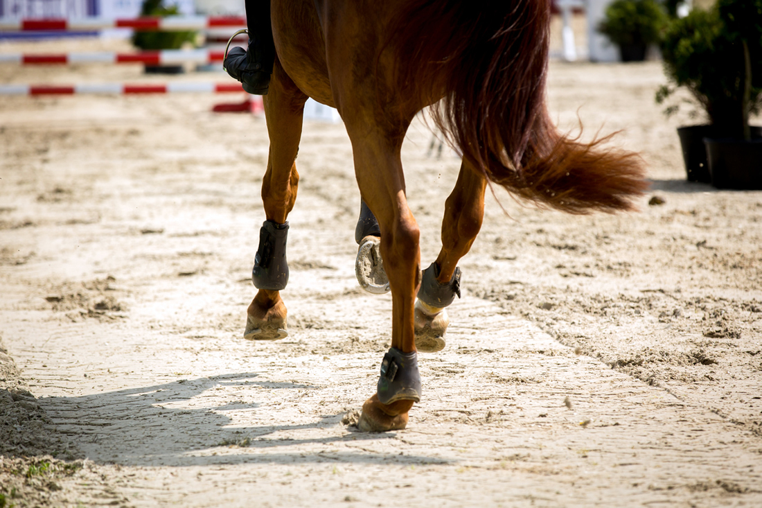
Bone spavin, also known as Degenerative Joint Disease (DJD) of the hock (tarsus), is a common cause of equine lameness.
It is caused by repeated concussion and rotational forces on the hock joint, as well as excessive forces on the adjoining ligaments.
As a wear and tear condition, bone spavin is characterized by narrowed joint spaces and bone spurs. [1][2] It is frequently bilateral and accompanied by lameness, with a decreased range of motion. [1]
It is mostly seen in mature equine athletes participating in sports that involve: [1][2][3]
- A lot of hock flexion, such as dressage
- Repeated jarring, such as jumping
- Rotational compressive forces, such as reining
- Repeated cyclical concussion, such as Thoroughbred or Standardbred racing
Bone Spavin in Horses
Two major factors lead to the development of bone spavin in horses:
- Cartilage compression affecting the distal tarsal bones: the central and third tarsals. Known as ‘cuboid’ bones, these are stacked one above the other. With continued, repetitive compression, the cartilage degenerates and becomes crushed, leading to narrowed joint spaces. These then fill with bone.
- Uneven loading leads to excessive compression of the cartilage and underlying bone on one side of the hock, with strain on the soft tissue structures located on the other side. This can lead to remodelling and the development of exostoses (bone spurs) which are outgrowths of bone. Pressure on the soft tissue structures can also lead to exostoses. [2]
 Illustration: Dr. Ana Mesa, PhD
 Conformational Factors
Additionally, sickle hocks and cow hocks are conformational factors that predispose to bone spavin and degenerative joint disease. [1][2][3]
Sickle-hocked horses have excessive angulation in the joints and stand with the hind feet well forwards, which creates excessive pressure down the plantar aspect (back) of the tarsus.
Cow-hocked horses have hocks that angle inwards and feet that angle outwards, leading to added pressure on the medial aspect of the tarsus.
Tarsal Joint Disease
The hock consists of five joints:
- The tarsocrural joint (also known as the tibiotarsal) articulates the tibia and the talus.
- The centrotarsal joint (proximal intertarsal) articulates the tarsus and central tarsal bone.
- The distal intertarsal joint articulates the central and third tarsal bones.
- The tarsometatarsal joint articulates the lowest hock bone and the third metatarsal (cannon).
- The talocalcaneal joint articulates the talus and the calcaneal bone (which forms the point of hock).
Most of the hocks’ movement occurs in the tarsocrural joint, while the other joints are almost immobile. Synovial effusion is often seen around the tarsocrural joint, if the horse is developing osteoarthritis, but not around the central tarsal and distal intertarsal.
Location-wise, degenerative joint disease mostly involves the lower joints of the hock. Sometimes it affects the proximal joints, particularly the tarsocrural joint. [1]
Types of Hock Spavin
Different types of hock spavin may develop, including:
- True bone spavin involves the radiographically visible osteoarthritis of the distal tarsal joints.
- Blind or occult spavin is not radiographically visible, although lesions may be present in the articular cartilage (this is a post mortem finding).
- Blood spavin indicates a significant swelling that causes distension of the saphenous vein where it runs over the joints.
- High spavin is osteoarthritis in the proximal tarsal joints. [1]
- In young horses, ‘juvenile spavin‘ is often associated with subchondral bone issues, such as osteochondrosis. [2]
Metabolic bone disease may also contribute to DJD, due to mineral imbalances or deficiencies, protein excess or deficiency.
Endocrine imbalances linked to hypothyroidism or gestational immaturity can also lead to incomplete ossification of the central and third tarsal bones in young foals. This can lead to collapse or wedging and subsequent abnormal loading. [1][2][4]
Bone spavin may also be secondary to direct trauma, or damage affecting the hock’s ligaments or other soft tissue structures, with inflammation leading to secondary cartilage damage. [3]
What's your top priority with your horse's health?
What's your top priority with your horse's health?
Pathophysiology
Bone spavin starts to develop on the dorsomedial (inner, frontal) aspects of the hock joints, progressing primarily to the front. [3]
Early radiographic changes may show cyst formation involving the adjacent subchondral bone. As this progresses, irregular atrophy occurs in the subchondral bone, which makes the joint spaces appear wider.
A local periosteal reaction then occurs. This involves the cells of the periosteum – a membrane covering the bone – slowly forming bone. This progresses at different speeds depending on the individual – in some, ankylosis (bony fusion) between the third and central tarsal can occur within a year. At this point, the lameness is resolved. [1]
With computed tomography imaging (CT scans), it has become apparent that multiple pathologies can be present in different tarsal joints at the same time as radiographically identifiable osteoarthritis, including: [5]
- Subchondral cyst-like lesions
- Osteolytic lesions
- Sclerosis
- Lesions in the tarsometatarsal interosseous ligament
Changes in subchondral bone thickness have also been identified in horses with hock pain and radiographic changes. [6]
Signs & Symptoms
Horses with bone spavin usually become gradually lame over a period of time, often showing a mild lameness that may shift from one leg to the other.
In mild cases, horses can warm up out of the lameness. However, the lameness may become more consistent over weeks and months, and the horse shows a growing reluctance to work.
The horse may be reluctant to pivot or circle in the direction of the most affected limb, with a tendency to place greater weight on the least affected limb.
The lameness may improve with medication, only to worsen once the drugs are stopped. In severe cases, the horse may get worse as exercise continues.
Lameness generally worsens with work over a few days, then resolves with rest. [1][2][3]
Changes in Gait
The lameness is commonly seen as a ‘hip hike‘. During the cranial phase of the most affected limb, the sounder limb pushes both hips upwards so that the less sound limb can swing forward with reduced joint flexion.
The pain caused by flexion leads to a lower foot flight arc and a shorter cranial phase to the stride, with a tendency to land toe-first. This causes the toe to become worn on the most affected side, or both sides if the condition is bilateral. [1][2]
The foot or shoe may also show more wear on the outside, as the horse is trying to avoid pressure on the medial aspect of the joint. [1]
Sometimes, the horse can be observed lifting its hind feet and flexing its limbs while at rest. [1]
Bone Remodelling
As bone spavin mostly develops in the medial aspect of the hock, this may be where a bony enlargement is seen on the lower hock. This may be harder to identify when the horse is a large-boned type.
With the distal tarsal joints affected, there is not usually any effusion. If bog spavin (swelling of the tibiotarsal joint) is also present, the tarsocrural joint may be involved, or there is a further problem. [2]
Diagnosis
Consult with your veterinarian if your horse is showing signs of lameness. Diagnosis of bone spavin may be based on:
- Presence of lameness with lowered foot flight, toe wear, and possibly outside wear on hoof or shoe
- Marked positive response to flexion test
- Nerve blocks
Visual Assessment
Caution is needed when assessing the signs of lameness, as hock and stifle issues can present similarly.
Enlargement of the head of the second metatarsal bone (the medial splint) may give the visual appearance of a bone spavin, so further palpation or radiography is required.
The hocks must be assessed laterally and from in front for asymmetrical development and swelling. However, this is not so reliable when bilateral bone spavins are present. [1][3]
Flexion Test
The initial veterinary check for bone spavin is the flexion test.
To assess for hock lameness, the veterinarian lifts the foot to sharply flex the hock for one to two minutes, before immediately trotting the horse. If the horse has hock pain, it will take a few steps showing increased lameness.
It is rare to get a false positive with this test. Both hindlimbs are usually tested, to compare limbs in the case of unilateral lameness, or to identify bilateral lameness. [1]
The veterinarian may apply firm finger pressure over the medial aspect of the lower tarsal joints to accentuate the pain response in cases of DJD. [2][3]
There is some discussion about the reliability of this test, as other conditions can elicit positive results.
Diagnostic Analgesia
Diagnostic analgesia can also help localize hock pain. In this procedure, the caudal tibial and deep peroneal nerves are blocked with local anesthetic.
This is a reasonably accurate diagnostic method, but it will not identify exactly which tarsal joints are affected. [1] Intra-articular anesthesia into the individual distal joints will help to localize the problem more accurately.
Caution is needed with this diagnostic approach as the joint spaces communicate, and the anesthetic may travel from the tarsometatarsal joint to the centrodistal and tarsocrural joints.
Additionally, if the horse has subchondral bone pain or extensive cartilage pathology, these blocks may not block the pain, giving a false negative for DJD. [3]
Radiographs
Radiographs (x-rays) are essential for an accurate diagnosis of bone spavin. Images usually show the involvement of:
- The third metatarsal (medial aspect of proximal end)
- Central and third tarsal bones (medial aspect)
- Centrodistal joint (central tarsal, third tarsal) – ankylosis (fusion)
- Tarsometarsal joint (third tarsal, third metatarsal) – ankylosis (fusion)
With at least five different views taken, radiographs are examined for:
- Narrowing of joint spaces
- Bone spurs / osteophyte formation
- Sclerosis of the bone
- Fusion (ankylosis), possibly with a bony callus bridging the joint
- Collapse of the central and third tarsal bones
- Fracturing of the distal bones
- Cysts or other lesions in the subchondral bone might indicate metaplasia and degeneration of the tissue [1]
Other Methods
Ultrasound may also be used in the evaluation of soft tissue around the affected joint, in conjunction with radiography.
Magnetic resonance imaging (MRI) has also been successfully used to identify multiple lesion sites in the hock. [6][7] Nuclear scintigraphy is used in some complex cases. [8]

Treatment
Consult with your veterinarian to determine the best treatment protocol for your horse.
The changes associated with bone spavin are irreversible, so treatment is based on managing the condition to improve comfort and mobility. Therapeutic targets include:
- Providing analgesia (pain relief)
- Controlling inflammation
- Limiting damage to articular tissues
- Promoting the healing of damaged cartilage
This may involve non-steroidal anti-inflammatory drugs (NSAIDs) and/or corticosteroids to address pain, inflammation and lameness while controlling the progression of the condition.
These are often combined with oral nutraceuticals in a management program. [2][3]
Treatment Considerations
Treatment decisions are based on:
- The involved joint (or known to be involved)
- The stage of osteoarthritis
- The horse’s age
- Intended use for the horse
- Cost considerations
- Perceived likelihood of improvement
- Competition regulations (ie. use of controlled drugs) [3][9]
Medications
1) Non Steroidal Anti Inflammatory Drugs (NSAIDs)
NSAIDs are the frontline drug most commonly used to manage bone spavin, being cost-effective in reducing inflammation and lameness in many horses.
Long-term use can be counterproductive, as they lead to further degradation due to suppression of chondrocyte metabolism and inhibition of collagen and proteoglycan synthesis. [10]
Phenylbutazone (‘bute’) and firocoxib (Equioxx®) are the most commonly used NSAIDs. Disadvantages may present over time, depending on the dose and duration, in the form of gastrointestinal ulcers and renal papillary necrosis.
Right dorsal colitis (hindgut ulceration) has also been documented as a complication. [9]
2) Pentosan
Pentosan is the popular name for pentosan polysulfate, a polysulphated glycosaminoglycans (PSGAGs).
Injected intramuscularly, PSGAGs promote chondrocyte (cartilage cell) activity while inhibiting the negative effect of cytokines or prostaglandins on cartilage.
The exact mechanism is unknown, although one theory is that PSGAGs bond with fibronectin and collagen fibres in articular cartilage. There is also a low-level anti-inflammatory effect.
Pentosan can be administered into the joints, but severe infections have been reported so this is generally avoided. [3][9]
3) Bisphosphonates
Bisphosphonates such as tiludronic acid and clodronic acid slow down bone resorption and remodelling and may also be mild anti-inflammatories.
Tiludronate is licensed for use with bone spavin. Tildren is used widely. [3]
4) Intra-articular medications
Medications injected into the distal tarsal joints aim to reduce inflammation and pain associated with bone spavins.
Corticosteroids
Injected into the joints, corticosteroids are the most potent anti-inflammatory agents used to treat DJD. Corticosteroids such as methylprednisolone are used in the centrodistal joint, while triamcinolone is often used in the high-motion tarsocrural joint.
Accuracy of injection is sometimes an issue, particularly with the centrodistal joint; radiographs may be needed to assist needle placement. [3][11]
The disadvantage is that at high concentrations, corticosteroids can induce deterioration of the articular cartilage. At the same time, other steroids are known to have chondroprotective properties.
There is a risk of septic arthritis associated with the injections. The gravest risk associated with glucocorticosteroids is that of acute laminitis, with circumstantial evidence suggesting that very high doses and individual sensitivity might lead to this complication. [3][9]
Hyaluronic Acid (HA)
Hyaluronic acid is a component of both synovial fluid and articular cartilage. Injected into the joint, HA brings viscoelasticity to synovial fluid and lubrication to the synovial membrane and articular cartilage.
Beneficial effects of HA on cartilage metabolism have been demonstrated in horses after both intra-articular and intravenous administration. [9]
The molecular weight of HA has been shown to influence effectiveness: high and intermediate molecular weight products are more effective in the healing process, while very low molecular weight products can actually cause inflammation. [12]
Hyaluronic acid is also available as an oral joint supplement.
Polyacrylamide gel
Polyacrylamide gels such as Arthropen are a recent addition to the list of intra-articular medications.
Arthropen has been found in some studies to alleviate lameness and joint effusion, but there is currently insufficient evidence to show it is more effective than corticosteroids. [13]
Chemical arthrodesis
Chemical arthrodesis involves the injection of ethyl alcohol into the distal joints to prompt ankylosis (fusion) of the tarsometatarsal joint.
If osteoarthritis is already advanced and the distal joints are already ankylosing, this can accelerate full fusion and reduce inflammation and lameness. [1][3]
Stem cells
Injected into the joints, mesenchymal stem cells (MSCs) can improve cartilage healing and may reduce the symptoms of DJD. However, they will not prevent further degradation of the joint. [14]
5) Oral Nutraceuticals
This term is reserved for oral supplements that are neither pharmaceutical nor nutritional in nature.
Glucosamine & Chondroitin
Glucosamine-chondroitin sulfate is one such product that is administered extensively in cases of bone spavin due to its purported anti-inflammatory properties.
However, efficacy studies produce mixed results. [10] This inconsistency may be explained by variation in the quality of commercial preparations as well as the methodologies used, namely with regard to dosage.
Glucosamine salts and chondroitin sulphate are extracted from animal products or provided in a synthetic form.
- The glucosamine used in supplements is laboratory made or is derived from shellfish shells, such as the green lipped mussel. Forms include glucosamine sulfate, glucosamine hydrochloride, and N-acetyl glucosamine. [15]
- Chondroitin is made from bovine (cow) or shark cartilage. Chondroitin sulfate can also be synthetically produced.
Delivered orally in feed, glucosamine is well absorbed. Debate continues on the enteral absorption and bioavailability of chondroitin sulphate, which is far lower. [9]
Omega-3 Fatty Acids
Omega-3 fatty acids have anti-inflammatory effects in the body to reduce inflammation and support tissue repair. In particular, the omega-3s DHA (docosahexanoic acid) and EPA (eicosapentanoic acid) are the active omega-3s that have anti-inflammatory effects in the body.
DHA and/or EPA are derived from marine sources such as fish oil and microalgae. These can be safely fed to horses to provide anti-inflammatory benefits. Regarding joint health, EPA and DHA supplementation in horses has been shown to: [19][20][21][22]
- Increase stride length suggesting improved joint comfort
- Reduced pro-inflammatory markers
- Altered synovial fluid composition
Mad Barn’s w-3 oil provides 1,500 mg microalgal DHA per typical 100 ml serving.

Methylsulfonylmethane (MSM)
MSM is a naturally available form of sulfur found in plants. It can also be added to equine diets as a supplement to reduce exercise-induced inflammation. [17]
It may also protect against loss of cartilage and reduce inflammation in joints. [18]
Mad Barn’s pure MSM powder is a cost-effective option for adding a bioavailable form of sulfur to your horse’s diet.
Surgical Options
Joint lavage is a potential tool for managing joint disease in horses. This procedure removes degenerative debris and inflammatory cells from the joint. [16]
A more invasive technique for fusing the tarsometatarsal and centrodistal joints involves drilling the joint; this is generally successful. [1]
Cunean tenectomy is usually performed when other measures have failed to achieve improvement. This involves removing a small section of the cunean tendon, which decreases the rotational forces affecting the joint.
However, this procedure is seen as appropriate only when new bone forms under the tendon and causes pain. [1][16]
Laser treatment (photobiomodulation) has produced successful outcomes for DJD of the centrotarsal and tarsometatarsal joints when applied directly into the joint in surgical conditions.
It is possible that the pain sensation is eliminated with the destruction of nerve endings, as no fusion occurs. [1]
Therapeutic Approaches
Other approaches utilizing veterinary therapeutic devices are also used for bone spavin. These include shockwave therapy (SWT), which is reported to have an analgesic effect.
Cryotherapy (cold therapy) is another approach used for managing inflammation. Low-level laser therapy also addresses inflammation and pain.
Hydrotherapy in the form of swimming in purpose-built pools or in natural bodies of water has also been recommended. [16]
Hoof balancing is an important aspect of bone spavin management and supporting joint health. As well as reducing rotational and shear forces by improving breakover, bringing the toe back can help to improve stability. [1][3]
Prognosis
The prognosis for bone spavin depends upon:
- The number of affected joints. If there are bony changes in the tarsocrural joint and the distal tarsal joints, the prognosis is usually poor.
- The severity of the bony changes. If there is ankylosis or fusion of the intertarsal and/or tarsometatarsal joints, either through natural progression or arthrodesis of the joint, the prognosis is a rideable horse. There will usually be no lameness once warmed up. The horse may still have a lower foot flight arc.
- The horse’s age and speed of progression. If slower, a milder DJD presentation will be more manageable for longer.
- Previous or intended athletic purpose of the horse. A return to higher level athletic performance is unlikely. However, with appropriate management, many horses can continue in light riding for years. [1][2]
If your horse has new or worsening lameness, consult with your veterinarian to get an accurate diagnosis. An equine nutritionist can also help you formulate a feeding program to support bone and joint health.
Frequently Asked Questions
Here are some frequently asked questions about bone spavin in horses:
Bone spavin refers to degenerative joint disease affecting the lower hock joints. Repeated compression and rotational forces damage cartilage, narrow the joint spaces, and trigger bony changes that limit mobility. Horses often develop gradual lameness, stiffness, or reluctance to work as inflammation and joint wear progress over time.
Early signs often include subtle stiffness, shorter strides, or a noticeable hip-hike as the horse avoids flexing the affected joint. Some horses warm up out of the lameness briefly before worsening with more work. Toe wear, reluctance to circle, difficulty engaging the hind end, or shifting lameness between limbs can also signal developing hock pain.
Horses in disciplines that place repeated stress on the hock—such as dressage, jumping, reining, and racing—face a higher risk. Conformational traits like cow hocks or sickle hocks increase uneven loading and accelerate joint wear. Mature athletic horses, individuals with prior hock injuries, and those exposed to chronic concussion are also more prone to degenerative changes.
Management focuses on easing pain, controlling inflammation, and slowing joint deterioration. Many horses improve with balanced hoof care, appropriate exercise, weight control, and veterinary-guided therapies such as NSAIDs, joint injections, or supportive supplements. Consistent farriery that improves breakover reduces torque on the hock, and thoughtful conditioning programs help many horses stay comfortable in long-term work.
Summary
Bone spavin is a degenerative joint disease of the hock in horses. It describes wear of the lower hock joints caused by long-term mechanical stress.
- Cartilage deterioration narrows joint spaces and leads to new bone growth
- Uneven loading increases strain on the medial side of the hock
- Joint changes gradually reduce flexion and alter normal stride mechanics
- Progressive stiffness and intermittent lameness are common clinical signs
- Work demands, footing, and conformation can influence disease progression
References
- Baxter G. Adams' Lameness in Horses. 4th ed. Wiley Blackwell; 2011.
- King C, Mansmann, R. Lameness. 2005 ed. Guilford, Connecticut: The Lyons Press; 1995.
- Ross M, Dyson, S. Diagnosis and Management of Lameness in the Horse. St Louis, Missouri: Elsevier Saunders; 2011.
- Sedrish SA, Moore RM. Diagnosis and management of incomplete ossification of the cuboidal bones in foals. Equine Practice. 1997.
- Raes E, Bergman HJ, Van Ryssen B, Vanderperren K, Stock E, Saunders JH. Computed tomographic features of lesions detected in horses with tarsal lameness. Equine Veterinary Journal. 2014. View Summary
- Branch MV, Murray RC, Dyson SJ, Goodship AE. Alteration of distal tarsal subchondral bone thickness pattern in horses with tarsal pain. Equine veterinary journal. 2007. View Summary
- Barrett MF, Selberg KT, Johnson SA, Hersman J, Frisbie DD. High field magnetic resonance imaging contributes to diagnosis of equine distal tarsus and proximal metatarsus lesions: 103 horses.Veterinary Radiology & Ultrasound. 2018. View Summary
- McIlwraith CW, Kawcak CE, Frisbie DD, et al. Biomarkers for equine joint injury and osteoarthritis: Biomarkers for Equine Traumatic Arthritis and Osteoarthritis. Journal of Orthopaedic Research. 2018.
- Bolt D. Equine degenerative joint disease – medical treatment and management. Vet Times. 2013. Accessed December 5, 2022.
- Clayton H, et al. Double-Blind Study of the Effects of an Oral Supplement Intended to Support Joint Health in Horses with Tarsal Degenerative Joint Disease. American Association of Equine Practitioners. 2002.
- Seabaugh K.A. et al. Clinical study evaluating the accuracy of injecting the distal tarsal joints in the horse. Equine Veterinary Journal. 2017. View Summary
- Neuenschwander, H.M. et al. Hyaluronic acid has chondroprotective and joint-preserving effects on LPS-induced synovitis in horses. Journal of Veterinary Science. 2019. View Summary
- Bowkett-Pritchard, C.Equine osteoarthritis: is intra-articular polyacrylamide gel more effective than corticosteroid in improving lameness? Veterinary Evidence. 2022.
- Broeckx S, et al. Equine Allogeneic Chondrogenic Induced Mesenchymal Stem Cells Are an Effective Treatment for Degenerative Joint Disease in Horses. Stem Cells and Development. 2019. View Summary
- Staff MC. Glucosamine. Mayo Clinic. 2020.
- Auer JA, Fackelman GE. Treatment of Degenerative Joint Disease of the Horse: A Review and Commentary. Veterinary Surgery. 1981.
- Butawan, M. et al. Methylsulfonylmethane: Applications and safety of a novel dietary supplement. Nutrients. 2017.
- Marañón, G. et al. The effect of methylsulphonylmethane supplementation on biomarkers of oxidative stress in sport horses following jumping exercise. Acta Vet Scand. 2008.
- Ross-Jones, T. et al. Effects of omega-3 long chain polyunsaturated fatty acid supplementation on equine synovial fluid fatty acid composition and prostaglandin E2. J Equine Vet Sci. 2014.
- Woodward, AD. et al. Supplementation of dietary long-chain polyunsaturated omega-3 fatty acids high in docosahexaenoic acid (DHA) increases plasma DHA concentration and may increase trot stride lengths in horses. Equine Comp Exerc Physiol. 2007.
- Manhart, DR. et al. Markers of inflammation in arthritic horses fed omega-3 fatty acids. Prof Anim Sci. 2009.
- Vanderweerd, JM. et al. Systematic review of efficacy of nutraceuticals to alleviate clinical signs of osteoarthritis. J Vet Intern Med. 2012. View Summary


