
 English

Thoroughpin refers to a soft, fluid-filled swelling that develops around the back of a horse’s hock. It occurs when excess fluid collects within the tarsal sheath, a structure surrounding a tendon that passes along the back of the joint.
In many horses, thoroughpin is painless and does not cause lameness. The swelling may persist without affecting movement or athletic ability, making it primarily a cosmetic concern. However, a similar swelling can also develop when the tarsal sheath is affected by injury, inflammation, infection, or abnormalities involving nearby tissues or bone.
Distinguishing uncomplicated thoroughpin from a more significant problem is important for timely diagnosis and treatment, particularly when swelling appears suddenly or occurs with heat, pain, lameness, or a wound near the hock.
Understanding how thoroughpin develops, what other hock swellings can resemble it, and when veterinary evaluation is warranted can help ensure that underlying problems are identified and treated appropriately.
Thoroughpin in Horses
Thoroughpin is a soft, fluid-filled swelling around the back of a horse’s hock caused by excess fluid within the tarsal sheath.
The swelling may be visible on both the inside and outside of the hock. [1][2][3]
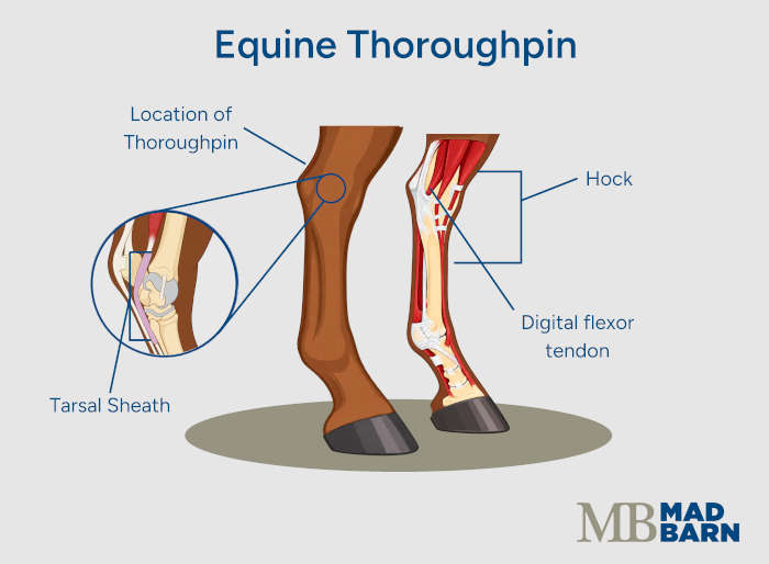
The tarsal sheath is a thin, fluid-filled structure around a tendon that passes along the back of the hock. The tendon, called the lateral digital flexor tendon, moves within this sheath as the horse bends and straightens the leg. A small amount of fluid inside the sheath helps the tendon move smoothly. [1][2][4]
The tarsal sheath extends above and below the hock. When extra fluid collects inside it, the sheath stretches and creates the soft swelling known as thoroughpin. [1][4]
Thoroughpin can occur without an identifiable injury or disease, making it idiopathic. This form of thoroughpin is generally painless and not associated with lameness.
In these cases, the swelling is primarily a cosmetic concern rather than a problem affecting the horse’s comfort or movement. This painless form without an identified underlying problem is referred to as “uncomplicated thoroughpin.” [1][2][3]
In some horses, swelling of the tarsal sheath develops because of an underlying problem such as injury, inflammation, or infection. These cases are more likely to cause pain or lameness and may require treatment. [1][5][6]
When owners first notice swelling around their horse’s hock, they should check the area for heat, pain, and wounds, and monitor their horse for lameness or difficulty moving the hock. Horses with painful or inflamed thoroughpin require rest and limited exercise until the underlying cause is determined. [1][3]
What's your top priority with your horse's health?

What's your top priority with your horse's health?
Thoroughpin vs. Other Types of Hock Swelling
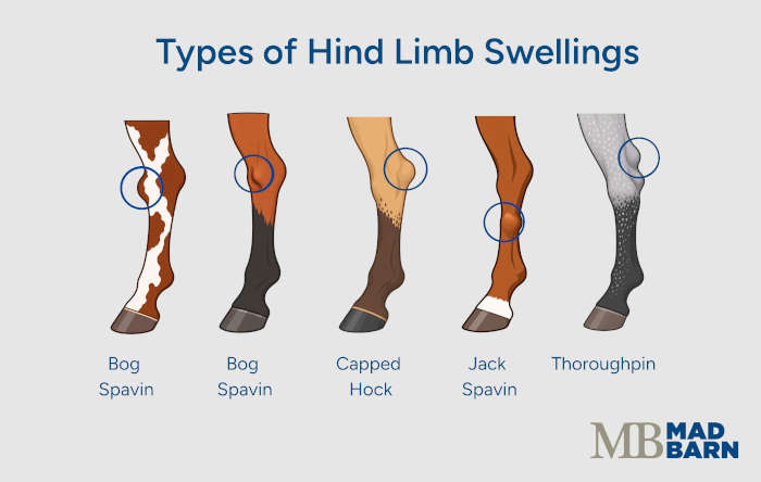
Several conditions can cause swelling around a horse’s hock. The location of the swelling and the structure containing the extra fluid help distinguish thoroughpin from other types of hock swelling. [1]
Thoroughpin vs. Bog Spavin
Thoroughpin and bog spavin both cause soft, fluid-filled swelling around the hock, but they involve different structures. Thoroughpin develops when fluid builds up within the tarsal sheath, while bog spavin is caused by excess fluid within the main hock joint. [1]
Thoroughpin is most noticeable at the back of the hock and may produce swelling on both the inside and outside. [1] Bog spavin is usually most noticeable toward the front and inside of the hock, although swelling can also appear toward the back. [1]
Other Hock Swellings
Capped hock causes swelling directly over the point of the horse’s hock. It commonly develops after repeated pressure or trauma to this area, such as kicking a stall wall. [1]
False thoroughpin is a fluid-filled swelling that looks similar to thoroughpin but does not involve simple fluid buildup within the tarsal sheath. It may form as a separate fluid-filled sac or when fluid pushes through a small defect in the wall of the tarsal sheath. [1][7]
Since these swellings can look similar from the outside, their appearance alone may not always be enough to identify the cause. [1]
Table 1. Thoroughpin vs. bog spavin and other hock swellings
| Type of Swelling | Location | Structure Involved | Clinical Signs |
|---|---|---|---|
| Thoroughpin |
|
|
|
| Bog spavin |
|
|
|
| Capped hock |
|
|
|
| False thoroughpin |
|
|
|
Signs of Thoroughpin in Horses
The main symptom of thoroughpin in horses is a soft swelling at the back of the hock. The swelling may be visible on both sides of the hock and can vary in size. [1]
With uncomplicated thoroughpin, the swelling is usually soft, cool, and painless, and the horse is not lame. The swelling may remain visible or come and go over time. [1][2]
When thoroughpin is related to an injury or inflammation, additional symptoms may include: [1][6]
- Warmth around the swelling
- Pain when the area is touched
- Lameness
- Pain when the hock is bent
- Reduced movement of the hock
Long-standing cases associated with injury may feel firmer as changes develop within and around the tarsal sheath. Pain and lameness vary considerably and do not always reflect how severe the underlying problem is. [1]
When to Call the Veterinarian
Veterinary evaluation is the only way to find out if the swelling is uncomplicated thoroughpin or related to an injury, inflammation, infection, or another type of hock swelling. [1][3]
Prompt veterinary attention is particularly important when there is: [1][3]
- Lameness
- Heat
- Pain
- A wound near the hock
- Rapidly increasing swelling
Owners should treat a wound near the tarsal sheath as potentially serious because infection within the sheath can cause significant damage if treatment is delayed. [1][3] Once uncomplicated thoroughpin has been diagnosed and the horse is comfortable and sound, treatment or exercise restriction may not be necessary. [1]
Causes of Thoroughpin in Horses
Thoroughpin often develops without an obvious injury or disease, especially in young horses. In such cases, fluid simply builds up within the tarsal sheath, usually without causing pain or lameness. [1][2]
Thoroughpin can also develop when the tarsal sheath or nearby tissues are injured or inflamed. Causes include: [1][2][5][6]
- A kick or other direct injury to the hock
- A sprain or other soft tissue injury
- Damage to the tendon that runs through the tarsal sheath
- A fracture, bone fragment, or abnormal bone growth next to the tarsal sheath
- Infection within the tarsal sheath
In these cases, damage or inflammation causes additional fluid to collect within the sheath. [5][6] Thoroughpin caused by an underlying injury or infection is more likely to occur with pain or lameness. [1][2]
Diagnosis of Thoroughpin
Veterinary investigation of thoroughpin is warranted when the swelling: [1][3]
- Appears suddenly
- Becomes painful
- Causes lameness
- Develops after an injury or possible infection
The veterinarian’s goal is to determine whether the distension represents uncomplicated enlargement of the tarsal sheath or whether another problem affects the tendon, tendon sheath, nearby joints, or surrounding structures. [1][3]
The examination usually starts with inspection and palpation of the horse’s hock. The veterinarian assesses: [1][3]
- The size and location of the swelling
- Whether it feels soft or firm
- Whether pressure causes pain
- Whether the horse shows heat or lameness
Comparing both hind limbs can help determine whether the swelling is new, asymmetric, which raises the possibility of an underlying problem. [1][3]
Diagnostic tests may include: [1][3][8]
- Ultrasound
- X-rays
- Contrast X-rays
- Fluid sampling
- Tenoscopy
Ultrasound
Ultrasound is useful for examining the soft tissues within and around the tarsal sheath. It allows the veterinarian to assess tendon structure, sheath thickening, fluid accumulation, and other changes that could help explain persistent swelling or lameness.
X-rays
Standard radiographs (x-rays) provide information about the surrounding bones and joints. [1][3]
Contrast radiography involves injecting contrast material into the tendon sheath before taking x-ray images. This outlines the sheath and can reveal internal abnormalities that ordinary x-rays do not show clearly. [8]
Fluid Sampling
If infection or significant inflammation is a concern, the veterinarian can collect fluid from the tarsal sheath for laboratory analysis. Fluid characteristics and cell counts can help distinguish uncomplicated distension from more serious inflammatory or infectious disease. [1][3]
Tenoscopy
In some cases, veterinarians use tenoscopy to examine the inside of the tendon sheath directly with a small camera.
This provides a closer look at the tendons and inner surface of the sheath and is most useful when imaging suggests a structural lesion or when the cause of persistent swelling and lameness remains unclear. [1][3]

Treatment of Thoroughpin
Treatment of thoroughpin in horses depends on the cause of the swelling and whether the horse shows pain or lameness.
Uncomplicated thoroughpin that is soft, non-painful, and does not interfere with movement often requires no specific treatment. In these cases, veterinarians may recommend monitoring the swelling for changes rather than treating the appearance of the distended tarsal sheath itself. [1][3]
When inflammation of the tarsal sheath causes discomfort but diagnostic testing does not identify significant tendon or bone damage, treatment generally focuses on reducing inflammation and allowing the affected tissues to recover. This may include: [1][3]
- Rest or reduced exercise
- Anti-inflammatory medication
- Cold therapy
The treating veterinarian guides adjustments to the horse’s exercise program as swelling and lameness improve. Persistent distension does not always mean treatment has failed, particularly if the horse remains comfortable and sound. [1][3]
Surgical treatment is more likely when a structural problem inside the sheath continues to cause inflammation or lameness. Bone fragments, adhesions, or damaged tissue can irritate the tendon and the lining of the sheath, preventing the swelling from resolving with conservative treatment alone. [1][2][3]
Infection within the tarsal sheath requires prompt and aggressive treatment. Treatment may include antimicrobial therapy together with flushing or surgical cleaning of the sheath to remove infected fluid and damaged material. Delaying treatment increases the risk of permanent damage to the tendon and other structures within the sheath. [1][2][3]
Recovery & Prognosis
The outlook for uncomplicated thoroughpin is generally good. Horses without pain or lameness may remain fully active, although the swelling can persist and leave a cosmetic change in the appearance of the hock. [1][3]
When thoroughpin is caused by an injury, recovery depends on the amount of damage to the tendon, tarsal sheath, or nearby bone. Long-standing injuries are more likely to cause recurring swelling and lameness. [1][3]
The outlook is more guarded when there is severe tendon or bone damage or infection within the tarsal sheath. Prompt treatment of infection improves the chances of recovery. [1][2]
Prevention
There is no established way to prevent idiopathic thoroughpin because it can develop without an identifiable injury or disease. However, reducing injuries around the hock may help prevent some cases associated with trauma. [1][5][6]
Measures that may reduce a horse’s risk of thoroughpin include: [1][6]
- Maintaining safe stalls, fencing, and trailers to reduce the risk of hock injuries
- Reducing situations in which horses are likely to kick or be kicked
- Checking wounds around the hock promptly
- Seeking veterinary attention for wounds that may involve the tarsal sheath
Wounds around the tarsal sheath are particularly important because contamination can lead to infection within the sheath and damage to nearby tissues and bone. [1]
Pain, heat, lameness, rapidly increasing swelling, or a nearby wound can indicate an underlying problem involving the tarsal sheath or surrounding structures. Veterinary evaluation is important in these cases so that injuries, infection, and other causes of hock swelling can be identified and treated when necessary.
Frequently Asked Questions
Here are some frequently asked questions about thoroughpin in horses:
Thoroughpin in horses is a soft, fluid-filled swelling that develops at the back of the hock. It occurs when excess fluid collects within the tarsal sheath, which surrounds the lateral digital flexor tendon as it passes along the back of the joint. The swelling may be visible on both the inside and outside of the hock.
Thoroughpin in horses often develops without an identifiable injury or disease, particularly in young horses. It can also result from a direct injury, sprain, tendon damage, bone fragment, abnormal bone growth, or infection involving the tarsal sheath. Cases caused by an underlying injury or infection are more likely to involve pain, heat, or lameness.
Thoroughpin in horses is not usually serious when the swelling is cool and painless and the horse is not lame. This uncomplicated form may be primarily a cosmetic concern and might not affect movement or athletic activity. However, thoroughpin can also occur with tendon injuries, inflammation, bone abnormalities, or infection, so painful swelling or swelling accompanied by lameness requires veterinary evaluation.
The signs of uncomplicated thoroughpin include a soft, cool, painless swelling at the back of the hock without lameness. The swelling can vary in size, appear on both sides of the hock, and remain present or come and go over time. Warmth, pain, reduced hock movement, or lameness may indicate an underlying injury or inflammation instead.
Thoroughpin does not usually cause lameness when it develops without an underlying injury or disease. Lameness is more concerning for inflammation, tendon damage, a bone abnormality, infection, or another condition affecting the hock. The degree of lameness does not always reflect the severity of the underlying problem, so veterinary assessment may still be necessary.
A horse can often be ridden with thoroughpin once a veterinarian has identified the swelling as uncomplicated and the horse is comfortable and sound. Exercise may need to be restricted when the swelling is new, painful, warm, or associated with lameness until the underlying cause is determined. A horse with difficulty bending the hock or a wound near the swelling should not return to normal work without veterinary guidance.
Thoroughpin may go away on its own, but some horses continue to have a soft, painless swelling after other concerns have been ruled out. The swelling may also decrease and return over time. Persistent uncomplicated thoroughpin can leave a cosmetic change in the appearance of the hock without affecting the horse's comfort or ability to exercise.
The difference between thoroughpin and bog spavin is the structure in which excess fluid collects. Thoroughpin involves the tarsal sheath at the back of the hock, while bog spavin involves the main hock joint. Bog spavin is usually most noticeable toward the front and inside of the hock, but veterinary examination may be needed because these swellings can look similar.
Thoroughpin differs from capped hock in both its location and the structure involved. Thoroughpin is caused by fluid within the tarsal sheath and appears around the back of the hock, often on both sides. Capped hock produces swelling directly over the point of the hock and commonly develops after repeated pressure or trauma, such as kicking a stall wall.
Thoroughpin can occur in one hock or affect both hind legs. Swelling in only one hock raises the likelihood of an underlying problem, because idiopathic thoroughpin is usually bilateral, though it does not confirm one. Pain, heat, lameness, wounds, and sudden changes in the swelling are also important warning signs, and they matter more than laterality alone.
Thoroughpin is diagnosed by determining whether the swelling involves the tarsal sheath and whether an underlying injury, inflammation, or infection is present. A veterinarian may examine the hock and use ultrasound, radiographs, contrast radiographs, fluid sampling, or tenoscopy. These tests can help identify tendon damage, bone abnormalities, infection, and other conditions that may resemble uncomplicated thoroughpin.
A veterinarian should examine thoroughpin when the swelling is new, painful, warm, rapidly increasing, or accompanied by lameness or difficulty moving the hock. A wound near the swelling requires prompt attention because damage to the tarsal sheath can allow a serious infection to develop. Veterinary evaluation is also appropriate when the cause of the swelling is uncertain or its appearance changes.
Thoroughpin is treated according to its underlying cause and may not require treatment when it is painless and does not cause lameness. Inflammation without tendon or bone damage may be managed with rest, cold therapy, and veterinary-prescribed anti-inflammatory medication. Persistent cases involving damaged tissue, scar tissue, or bone fragments may require tenoscopy or another surgical procedure, while infection requires prompt treatment and may require surgery to clean and flush the sheath.
Thoroughpin cannot always be prevented because uncomplicated cases may develop without an identifiable injury or disease. The risk of trauma-related cases may be reduced by maintaining safe stalls, fencing, and trailers and limiting situations in which horses may kick or be kicked. Wounds around the hock should be checked promptly, and veterinary attention is important when a wound could involve the tarsal sheath.
Summary
Thoroughpin is a fluid-filled swelling of the tarsal sheath at the back of a horse's hock that may be cosmetic or associated with injury, inflammation, or infection.
- Uncomplicated thoroughpin is usually soft, cool, painless, and not associated with lameness.
- Heat, pain, lameness, rapid enlargement, or a nearby wound warrants prompt veterinary evaluation.
- Thoroughpin affects the tarsal sheath, while bog spavin involves the main hock joint and capped hock occurs over the point of the hock.
- Diagnostic testing may include ultrasound, X-rays, contrast radiography, fluid sampling, or tenoscopy.
- Treatment ranges from monitoring uncomplicated swelling to rest, surgery, or aggressive treatment for infection.
References
- Ross. M. W. and Dyson. S. J. Eds. Diagnosis and Management of Lameness in the Horse. 2nd ed. Elsevier/Saunders. 2011.
- Cauvin. E. R. J. et al. Endoscopic Examination of the Tarsal Sheath of the Lateral Digital Flexor Tendon in Horses. Equine Veterinary Journal. 1999.
- Baxter. G. M. Ed. Adams and Stashak's Lameness in Horses. 7th ed. Wiley-Blackwell. 2020.
- Auer. J. A. et al. Eds. Equine Surgery. 5th ed. Elsevier. 2019.
- Edwards. G. B. Changes in the Sustentaculum Tali Associated with Distension of the Tarsal Sheath (Thoroughpin). Equine Veterinary Journal. 1978.
- Dik. K. J. and Merkens. H. W. Unilateral Distension of the Tarsal Sheath in the Horse: A Report of 11 Cases. Equine Veterinary Journal. 1987.
- Minshall. G. J. and Wright. I. M. Synoviocoeles Associated with the Tarsal Sheath: Description of the Lesion and Treatment in 15 Horses. Equine Veterinary Journal. 2012.
- Hago. B. E. D. and Vaughan. L. C. Use of Contrast Radiography in the Investigation of Tenosynovitis and Bursitis in Horses. Equine Veterinary Journal. 1986.