English
Buttress foot, also known as pyramidal hoof disease, is a firm enlargement that develops at the front of a horse’s coronary band. It is associated with changes in the coffin bone or the nearby coffin joint. [1][2][3]
The condition can look different from one horse to another. Some horses are lame or sore around the coronary band, while others stabilize with little or no obvious discomfort. Changes in this area can also interfere with normal hoof growth, leading to ridges, uneven growth or distortion of the front hoof wall.
Common causes include fractures of the extensor process, previous trauma and arthritis in the coffin joint. Treatment and prognosis depend on the cause and the extent of coffin joint involvement.
Horses with new swelling around the coronary band, persistent foot pain or unexplained changes in hoof shape should be examined by a veterinarian.
What Is Buttress Foot in Horses?
Buttress foot is a descriptive term for enlargement at the front of the horse’s coronary band. This is caused by changes in the bone or joint underneath it.
The condition’s other name, pyramidal hoof disease, refers to the raised or pyramid-like shape that can develop at the front of the hoof. [1][3][4]
Rather than being a specific disease, buttress foot can result from several changes involving the coffin bone or coffin joint. [1][2]
The external appearance alone cannot show exactly what is happening inside the hoof. Identifying the cause is important because treatment for an old, stable bony enlargement will differ from treatment for a recent fracture or painful arthritis. [1][2][5]
Anatomy of the Coffin Bone & Coronary Band
The coffin bone, also called the distal phalanx, pedal bone, or P3, is the lowest bone in the horse’s limb. It sits inside the hoof capsule and forms part of the coffin joint. [1][2]
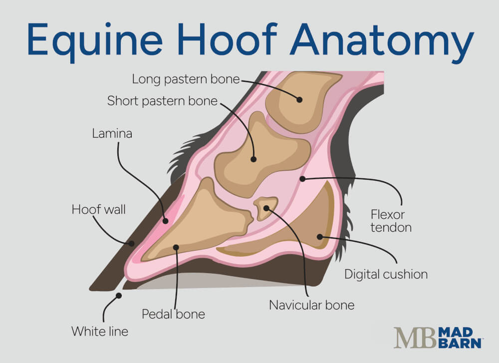 Illustration: Dr. Ana Mesa, PhD
 At the upper front portion of the coffin bone is a small projection called the extensor process. The digital extensor tendon attaches in this area and helps straighten the lower limb during movement. [4]
The extensor process lies close to both the coffin joint and the coronary band. A fracture or other change in this region can affect the nearby joint and create a visible bump at the top of the hoof. Changes in this area can also disrupt normal hoof growth, causing ridges or distortion in the hoof wall below the enlargement. [1]
Table 1. Buttress foot in horses at-a-glance
| Category | Key Information |
|---|---|
| Definition |
|
| Primary Cause |
|
| Common Causes |
|
| Onset |
|
| Symptoms |
|
| Severity |
|
| Treatment |
|
| Recovery Time |
|
| Prognosis |
|
| Prevention |
|
Signs of Buttress Foot in Horses
The most recognizable sign of buttress foot is a firm bump or swelling at the front of the coronary band, usually near the center of the hoof. [2]
Because the coronary band produces the hoof wall, changes in the tissues and bone underneath it can affect how the hoof grows. Over time, the hoof below the enlargement may develop an uneven, ridged or distorted appearance. [1]
Signs of buttress foot in horses include: [1][2][3][6]
- Firm swelling or a hard prominence at the front of the coronary band
- A raised or bulging appearance above the hoof wall
- Uneven or distorted hoof growth
- Ridges or changes in the shape of the front hoof wall
- Heat or tenderness around the coronary band
- Lameness ranging from mild to severe
- A shortened stride or reluctance to move freely
- Reduced athletic performance
The size of the enlargement does not always reflect the horse’s pain level. A horse with a large, longstanding bony prominence may be comfortable, while a smaller but more active problem involving the coffin joint may cause significant lameness. [1][2]
What's your top priority with your horse's health?

What's your top priority with your horse's health?
Recent vs. Longstanding Changes
Signs vary depending on whether the problem is recent or ongoing. With a recent injury, the area may develop heat and swelling, and the horse may show significant lameness. These early changes can subside before the underlying problem is diagnosed. [6]
Over time, the coronary band may become firm and thickened, while changes in hoof growth become more noticeable. The hoof wall may gradually develop the characteristic bulging or distorted appearance associated with buttress foot. [1][3]
When to Call the Veterinarian
Contact your veterinarian if you notice a new or enlarging bump around the coronary band, especially if the horse is lame or the area feels warm and painful.
Veterinary assessment is also recommended for: [1][2][7]
- Sudden or significant lameness
- Heat or increasing swelling around the coronary band
- Pain around the coronary band
- Recent trauma involving the hoof
- Progressive distortion of the hoof wall
- Persistent or recurring foot lameness
- A longstanding buttress foot that suddenly becomes painful or changes appearance
Several conditions can cause swelling or pain around the hoof. The outside appearance alone cannot identify which structures are involved. A veterinary examination and appropriate imaging can establish the cause and guide treatment.
Causes of Buttress Foot in Horses
Several problems can lead to buttress foot in equines. Some cases develop after an obvious injury, while others are linked to changes in the coffin joint that developed over time.
Extensor Process Fractures
Fractures of the extensor process can lead to buttress foot. These injuries occur after trauma to the hoof or when excessive force is placed on the front of the coffin bone. The fracture may involve a small piece of bone or a larger part of the extensor process. [5]
Because the extensor process forms part of the coffin joint, some fractures extend into the joint. This can lead to ongoing inflammation and increase the risk of arthritis over time. [5]
As the fracture heals, new bone can develop around the area. This bony growth may eventually result in the firm prominence known as buttress foot. [5]
Extensor Process Injury
Buttress foot can develop even when there is no primary fracture or arthritis within the coffin joint. In some cases, the injury affects the tendon and ligament fibers where they attach to the extensor process. [4]
Tearing or avulsion of these fibers can irritate the surface of the bone and trigger a repair response. As new bone accumulates at the attachment sites, the extensor process gradually enlarges and forms buttress foot. [4]
Osteoarthritis of the Coffin Joint
Arthritis in the coffin joint also contributes to buttress foot. Osteoarthritis damages the cartilage in a horse’s hoof, and the resulting inflammation may cause new bone to form around the edges of the joint. [2][7][8]
When new bone develops near the front of the coffin joint, it becomes visible or easy to feel at the coronary band. [7]
Horses with painful coffin joint arthritis may show: [7]
- Recurring lameness
- Stiffness
- Reduced performance
Signs may become more noticeable during exercise or when the horse works on firm or uneven ground. [7]
Trauma
Direct injury to the front of the hoof or coronary band can damage the underlying tissues and, in some cases, the coffin bone itself. [1][2][6]
A recent injury may cause swelling, pain and heat around the coronary band. If the bone or joint is involved, healing can leave behind permanent bony enlargement or changes in hoof growth. [1][2][6]
An injury to the coronary band does not always lead to buttress foot. The long-term outcome depends on which tissues were damaged and the severity of the injury. [1][2][5]
Developmental & Other Bony Abnormalities
Some horses develop bony fragments or irregularities around the extensor process, which may go unnoticed by owners until later in the horse’s life. These fragments can affect the coffin joint and may cause remodeling of the surrounding bone. [1][2][9]
A piece of bone found near the extensor process is not necessarily responsible for a horse’s lameness or hoof changes. Veterinarians compare imaging findings with the horse’s history, physical examination and response to diagnostic testing to determine whether the fragment is clinically significant. [1][2][10]
Risk Factors
The cause of buttress foot is usually more important than any single management factor. However, hoof conformation may influence the forces placed on structures around the extensor process. Horses with a very short toe and high heel are predisposed to pyramidal disease. [4]
Gait may also influence loading in this region. Some sources suggest that horses with a quick, short-stepping gait may have a higher risk of pyramidal disease, although research has not firmly established this relationship. [4]
A history of previous hoof injury, recurring foot soreness, or gradual changes in hoof shape can provide useful clues during a veterinary examination. These findings may help explain how the problem developed and identify factors contributing to ongoing strain. [1][2]
Diagnosis of Buttress Foot in Horses
Buttress foot is diagnosed based on the characteristic enlargement of the hoof, together with an assessment of the underlying cause and the extent of involvement of the internal hoof structures.
Because the visible change can result from several different disease processes, a complete diagnostic evaluation is needed to determine whether the condition is associated with pain or lameness and to identify the affected tissues. This may include: [1][2][6]
- A physical examination
- Lameness evaluation
- Nerve blocks
- Diagnostic imaging
Physical & Lameness Examination
During the physical examination, the veterinarian narrows their focus to the location of the buttress foot after completing a general assessment.
The veterinarian examines the hoof and coronary band for: [1][2][6][7]
- Swelling
- Heat
- Tenderness
- Uneven hoof growth
- Differences between the affected foot and the opposite limb
The veterinarian may observe the horse at the walk and trot, both in a straight line and on a circle. They may also walk the horse on different surfaces because some types of foot pain are easier to detect under certain conditions. Hoof testers can help identify areas of sensitivity within the foot, but they do not reveal the exact cause of the pain. [1][2]
Diagnostic Nerve Blocks
A veterinarian can use diagnostic nerve blocks to find the source of lameness when it is not obvious. [1][2]
This method temporarily numbs specific nerves or areas of the lower limb. The veterinarian then watches the horse move, and any improvement in their form helps narrow down where the pain is coming from. [1][2]
Nerve blocks locate pain but do not show what is physically wrong inside the hoof. Imaging is usually needed to identify the cause.
Radiographs
Radiographs are especially useful for evaluating buttress foot because they allow the veterinarian to see the coffin bone and coffin joint.
Several X-ray views may be needed to assess the area fully. Findings on x-ray may include: [1][2][5][10]
- Fractures of the extensor process
- Loose or separated bone fragments
- New bone formation
- Changes around the coffin joint
- Signs of arthritis
- Other abnormalities involving the coffin bone
The bump on the outside of the hoof does not always reflect how much damage is present underneath. Likewise, an old bone fragment on an X-ray may not be the reason a horse is currently lame. Radiographs are best interpreted alongside a thorough lameness examination. [1][2][5][10]
Other Diagnostic Imaging
Veterinarians may recommend additional imaging if radiographs do not fully explain the horse’s signs or if injury to nearby soft tissues is suspected. [1][2][10] Ultrasound can help evaluate some structures around the coronary band and lower limb. [1][2]
More advanced imaging, including magnetic resonance imaging (MRI) or computed tomography (CT), may provide more detailed information about structures inside the hoof in complicated cases. [10]
Treatment of Buttress Foot in Horses
There is no single definitive treatment for buttress foot. The appropriate treatment plan is based on the cause of the enlargement, how long it has been present, the severity of the lameness and whether the coffin joint is affected. [1][2][5][11]
Treatment of affected horses aims to: [1][2]
- Control pain and inflammation
- Allow injured tissues to heal
- Support comfortable movement
Rest & Exercise Management
Horses with a recent or painful injury may need a period of reduced activity to avoid placing additional stress on the affected area. [1][2][5]
The amount of rest depends on the diagnosis. A horse recovering from a fracture may need a different plan from one being managed for chronic coffin joint arthritis. [1][2][5]
Once the horse is comfortable and healing is progressing, the owner can gradually reintroduce exercise under veterinary guidance. A gradual approach is important, because returning to full work too quickly can place extra stress on healing tissues. A structured injury rehabilitation plan can help match activity to the stage of recovery. [1][2][5]
Pain & Inflammation Management
A veterinarian may prescribe non-steroidal anti-inflammatory drugs (NSAIDs) to reduce pain and inflammation. These medications can improve the horse’s comfort during recovery from a recent injury or help manage pain associated with chronic coffin joint disease.
Cold therapy may also help control pain, heat and swelling during the early stages of a recent injury. [1][2]
Pain medication and cold therapy can relieve symptoms, but they do not resolve the source of the condition. The veterinarian should reassess horses with persistent or worsening lameness to determine whether they require additional treatment. [1][2]
Hoof Care
Regular trimming and, when appropriate, supportive shoeing can help maintain hoof balance and improve comfort during recovery or long-term management. [1][2][3]
There is no universal trimming or shoeing approach that is suitable for every horse with buttress foot. The ideal plan depends on the horse’s hoof shape, conformation, diagnosis and workload. [1][2][3]
In many cases, collaboration between the veterinarian and farrier helps ensure that hoof care supports the affected area without adding unnecessary strain.
Treatment of Extensor Process Fractures
Some distal phalanx fractures can be managed conservatively with a period of rest, while certain extensor process fractures require surgical treatment. Treatment is guided by the size, location and age of the fracture, as well as whether it involves the coffin joint. [5][11]
Surgery is typically required to remove a bone fragment, particularly if the fragment continues to cause joint inflammation or lameness. The veterinarian can determine whether conservative treatment or surgery is more appropriate based on the horse’s X-rays and clinical signs. [9][11]
Coffin Joint Treatment
If inflammation or arthritis in the coffin joint is contributing to lameness, treatment may focus on reducing joint pain and keeping the horse comfortable. Depending on the horse, this may include: [1][2][8]
- Anti-inflammatory medications
- Joint injections
- Adjustments to exercise or hoof care
Established osteoarthritis in horses is irreversible. Long-term management focuses on reducing discomfort, supporting joint function and maintaining an appropriate level of activity. [1][2][8]
Recovery & Prognosis
The prognosis for recovery from buttress foot depends on the cause and the extent of coffin joint involvement. Horses with a small, stable problem and little joint damage may have a better outlook than horses with significant arthritis or ongoing joint pain. [5] The horse’s comfort and the joint involvement have more impact on prognosis than the appearance of the hoof.
The visible enlargement may remain even after the horse becomes comfortable, as bony prominences are permanent. [1][2] Changes in the hoof wall also take time to improve. Because new hoof grows down from the coronary band, damaged or distorted hoof may take several months to grow toward the ground. [1][7]
Some horses can return to their previous level of work, while others may need a lighter workload or ongoing management. [9]

Prevention
Not every case of buttress foot is preventable. Fractures can result from accidents, while some pieces of bone around the extensor process may have developmental origins. Coffin joint arthritis can also develop despite appropriate care.
Prevention focuses on reducing unnecessary stress and trauma to the hoof and identifying injuries before they lead to extensive bone or joint changes.
Maintain Balanced Hooves
Regular trimming and appropriate shoeing help maintain hoof balance and support even loading of the lower limb. The farrier should tailor hoof care to the horse’s: [1][2]
- Conformation
- Movement
- Workload
- Existing hoof shape
Horses with a short toe and high heel may place different forces on the extensor process and its tendon attachments. These horses may benefit from closer veterinary and farrier monitoring, although trimming or shoeing cannot eliminate every risk associated with conformation. [1][2][4]
Condition Horses Gradually
Sudden increases in exercise speed, duration or intensity can expose a horse’s unprepared bones and joints to greater loading. Horses returning to work after time off should follow a gradual conditioning program that allows these structures to adapt. [1][2]
Workload should also match the horse’s age, fitness, conformation, and previous injuries. Owners should reduce or stop exercise if a horse develops a shortened stride or reluctance to move forward. Ensuring horses have adequate rest and recovery time between training sessions also helps prevent over-exertion and reduces the chance of injury.
Provide Safe Footing & Surroundings
Safe, well-maintained footing helps reduce slipping and uneven loading. Owners should address deep holes, excessively hard ground, and slippery or irregular surfaces in riding and turnout areas promptly. [1][2][5]
Regularly checking a horse’s common areas for hazards also reduces the risk of other injuries.
Inspect the Hooves Regularly
Owners should examine and handle their horse’s feet routinely so they become familiar with each hoof’s normal shape and temperature.
Signs to watch for include: [1][2][3][6]
- New swelling or firmness around the coronary band
- Heat or tenderness
- Cuts or other injuries near the top of the hoof
- New ridges or uneven hoof growth
- Changes in stride or willingness to move
- Persistent or recurring foot lameness
Prompt veterinary assessment is particularly important after trauma or when swelling occurs with lameness. Radiographs can help identify an extensor process fracture, a separated piece of bone or coffin joint damage that may not be apparent from the outside. [1][2][5]
Frequently Asked Questions
Here are some frequently asked questions about buttress foot in horses:
Buttress foot in horses is a firm enlargement at the front of the coronary band caused by changes in the underlying coffin bone or coffin joint. It is a descriptive term rather than a single disease and can result from fractures, injuries or arthritis. New bone formation may create a raised appearance and interfere with normal hoof wall growth.
Buttress foot and pyramidal hoof disease are names for the same general condition. The term pyramidal hoof disease describes the raised or pyramid-like shape that can develop at the front of the coronary band. Both terms refer to an external enlargement associated with changes in the bone or joint underneath it.
Signs of buttress foot in horses commonly include a firm bump at the front of the coronary band and uneven, ridged or distorted hoof growth. A recent injury may also cause heat, tenderness, soft-tissue swelling and lameness. Longstanding cases may leave a hard prominence even when the horse is comfortable.
Buttress foot in horses can result from an extensor process fracture, injury where tendon or ligament fibres attach to the bone, coffin joint arthritis or direct trauma to the hoof. These problems can trigger new bone formation near the front of the coffin bone or coffin joint. Some bony irregularities may have developed earlier in the horse's life and may not be responsible for current lameness.
An extensor process fracture in horses is a break in the small projection at the upper front of the coffin bone. Because the extensor process forms part of the coffin joint, some fractures can cause persistent joint inflammation and increase the risk of arthritis. New bone that develops during healing may produce the characteristic enlargement at the coronary band.
Buttress foot can cause lameness, but a visible enlargement does not always mean the horse is in pain. Lameness depends on the underlying cause, the level of active inflammation and the extent of coffin joint involvement. A large, longstanding bump may cause little discomfort, while a smaller active injury can produce significant pain.
A horse with buttress foot may be ridden if the underlying condition is stable and the horse is comfortable, but suitability for work should be determined individually. Horses with active lameness, heat, tenderness or a recent injury should be evaluated by a veterinarian before continuing exercise. When work resumes, activity should be increased gradually according to the horse's diagnosis and recovery.
Buttress foot is diagnosed through the horse's history, a physical examination, a lameness assessment and diagnostic imaging. Radiographs can reveal extensor process fractures, bone fragments, new bone growth and arthritis in the coffin joint. Diagnostic nerve blocks may help locate the source of pain, while ultrasound, magnetic resonance imaging or computed tomography may be used in complicated cases.
Buttress foot can often be treated or managed, but there is no single treatment that is appropriate for every horse. The plan may include reduced activity, prescribed anti-inflammatory medication, rehabilitation and individualized hoof care. Some horses with painful extensor process fragments may require surgery, while those with established coffin joint arthritis may need long-term management.
Hoof care for a horse with buttress foot should focus on maintaining hoof balance and supporting comfortable movement. Regular trimming and, when appropriate, supportive shoeing may reduce unnecessary strain during recovery or long-term management. The best approach depends on the horse's hoof shape, conformation, diagnosis and workload, so coordination between the veterinarian and farrier is often beneficial.
Swelling from buttress foot may improve when it involves soft tissues affected by a recent injury. A hard bump can remain permanently once new bone has formed, even after inflammation and pain have resolved. Distorted hoof wall may also take several months to grow down from the coronary band.
A veterinarian should examine suspected buttress foot when a new or enlarging bump appears at the coronary band, particularly if the area is warm, swollen or painful. Sudden or persistent lameness, recent hoof trauma, progressive hoof wall distortion and recurring foot soreness also warrant assessment. A longstanding enlargement should be re-evaluated if it suddenly becomes painful or changes in appearance.
The prognosis for a horse with buttress foot depends primarily on the cause and the extent of coffin joint involvement. Horses with stable changes and little joint damage may return to their previous workload, while ongoing arthritis or joint pain can limit performance. The horse's comfort and joint health are generally more important to the outlook than the size or appearance of the enlargement.
Buttress foot in horses cannot always be prevented because accidental injuries and some extensor process abnormalities may occur despite good management. Regular hoof care, suitable conditioning and prompt attention to persistent foot pain can support lower-limb health. Frequent hoof inspections can also help owners detect coronary band injuries, new swelling and changes in hoof growth early.
Summary
Buttress foot is a firm enlargement at the front of a horse's coronary band that can result from injury, abnormal bone formation or disease affecting the coffin joint.
- Buttress foot can develop after extensor process fractures, trauma, attachment injuries, coffin joint arthritis or other bony abnormalities
- Affected horses may have coronary band swelling, hoof wall distortion, tenderness, a shortened stride or lameness
- Veterinarians assess whether the enlargement is painful and use imaging to identify changes in the coffin bone, coffin joint and nearby tissues
- Treatment depends on the cause and may include rest, anti-inflammatory medication, hoof care, joint treatment or surgery
- The enlargement may remain permanently, while prognosis depends mainly on coffin joint damage and ongoing pain
References
- Baxter. G. M. Ed. Adams and Stashak's Lameness in Horses. 7th ed. Wiley Blackwell. 2020.
- Ross. M. W. and Dyson. S. J. Eds. Diagnosis and Management of Lameness in the Horse. 2nd ed. Elsevier/Saunders. 2011.
- O'Grady. S. E. et al. A Glossary of Therapeutic Farriery Terms. Equine Veterinary Education. 2007.
- Rooney. J. R. Ringbone vs. Pyramidal Disease. Journal of Equine Veterinary Science. 1981.
- Rijkenhuizen. A. B. M. et al. Management and Outcome of Fractures of the Distal Phalanx: A Retrospective Study of 285 Horses With a Long-Term Outcome in 223 Cases. The Veterinary Journal. 2012.
- Reeks. H. C. Diseases of the Horse's Foot. Creative Media Partners, LLC. 2025.
- McAuliffe. S. B. Ed. Knottenbelt and Pascoe's Color Atlas of Diseases and Disorders of the Horse. 2nd ed. Saunders/Elsevier. 2014.
- Baker. M. E. et al. Investigation of MicroRNA Biomarkers in Equine Distal Interphalangeal Joint Osteoarthritis. International Journal of Molecular Sciences. 2022.
- Compagnie. E. et al. Arthroscopic Removal of Large Extensor Process Fragments in 18 Friesian Horses: Long-Term Clinical Outcome and Radiological Follow-Up of the Distal Interphalangeal Joint. Veterinary Surgery. 2016.
- Whitton. R. C. et al. The Diagnosis of Lameness Associated With Distal Limb Pathology in a Horse: A Comparison of Radiography, Computed Tomography and Magnetic Resonance Imaging. The Veterinary Journal. 1998.
- Crowe. O. M. et al. Long-Term Outcome After Arthroscopic Debridement of Distal Phalanx Extensor Process Fragmentation in 13 Horses. Veterinary Surgery. 2010.