English

Causes of lameness in horses range from common hoof problems to serious injuries involving joints, bones, tendons, ligaments, muscles, or the nervous system. Some causes appear suddenly, while others develop gradually and first show up as stiffness, reluctance to work, or a subtle decline in performance.
Lameness is not a disease itself. It is a clinical sign that something is affecting the horse’s normal movement. Pain is the most common reason horses become lame, but restricted movement, mechanical problems, neurological disorders, infection, and some systemic conditions can also change how a horse stands or moves.
Because the visible degree of lameness does not always match the seriousness of the underlying problem, owners should take both obvious and subtle gait changes seriously.
Keep reading to learn more about the common causes of lameness in horses, signs owners may notice, what can cause sudden lameness, and when veterinary care is needed to identify the source of the problem and prevent further injury.
Causes of Lameness in Horses
Lameness is one of the most common signs of pain or musculoskeletal injury in horses. It can appear as an obvious limp, but many cases begin with much subtler changes in stride, posture, willingness to move, or athletic performance. [1]
Lameness is not a condition itself, but a clinical sign that something is affecting the horse’s normal movement.
Pain is the most common cause of lameness, and may originate in the hoof, joints, bones, tendons, ligaments, muscles, or other supporting tissues. Restricted movement and disorders affecting the nervous or muscular systems can also alter the horse’s gait. [1]
How lame a horse appears does not always reflect the severity of the underlying problem. A hoof abscess can cause dramatic, non-weight-bearing lameness, while some stress fractures and soft-tissue injuries may initially cause only mild or intermittent changes in movement. [1][2]
Not all gait abnormalities are due to musculoskeletal pain. Other factors that impact the way a horse stands or moves include: [3]
- Infection
- Metabolic disease
- Restricted movement
- Disorders affecting the nervous or muscular systems
The source of lameness is not always obvious from the gait abnormality alone. Horses may alter their movement to reduce loading on a painful structure, shifting forces to other parts of the body and potentially contributing to secondary soreness.
In some cases, pain may also originate from more than one area, making the underlying cause more difficult to localize. [4]
Table 1. Overview of common causes of lameness in horses
| Source | Common causes | Possible signs | Important considerations |
|---|---|---|---|
| Hoof |
|
|
|
| Joints |
|
|
|
| Bone |
|
|
|
| Tendons and ligaments |
|
|
|
| Muscles, back, and pelvis |
|
|
|
| Neurological or mechanical |
|
|
|
| Trauma and wounds |
|
|
|
What's your top priority with your horse's health?

What's your top priority with your horse's health?
Hoof-Related Causes of Lameness in Horses
The hoof is a common source of lameness in horses. Within the rigid hoof capsule are several structures that can generate pain, including sensitive soft tissues, bone, joints, tendons, ligaments, blood vessels, and nerves. [5]
Because many of these structures are enclosed within the hoof capsule, significant pain may occur with few visible changes from the outside. As a result, hoof-related lameness can be severe even when swelling or other abnormalities higher in the limb are absent. [6][7]
Hoof Abscesses
A hoof abscess is a localized infection that develops when bacteria enter the hoof and become trapped within the hoof capsule. Entry may occur through a crack, white line defect, puncture, or damaged sole, although an obvious entry point is not always found. [7]
As infection and inflammation develop within the confined hoof capsule, increasing pressure can cause rapid-onset pain. Lameness may become severe or non-weight-bearing, and the affected hoof may feel warm with a stronger digital pulse. Visible swelling is often absent. [1][7]
Some abscesses eventually drain through the sole, heel bulbs, or coronary band, which typically relieves pressure. A veterinarian or farrier may use hoof testers and careful examination to help localize the painful area. [7][8]
If a suspected abscess does not follow the expected course, the affected hoof may require further veterinary investigation. Radiographs (x-rays) or other diagnostic tests can help rule out fractures, penetrating injuries, or other causes of hoof pain. [1][6][7]
Sole Bruising
A sole bruise occurs when sensitive tissues beneath the sole are damaged by excessive pressure or impact. Stones, hard or uneven ground, and repeated concussion can contribute, while horses with thin soles, flat feet, or inadequate hoof protection may be more susceptible. [9]
The sole may appear normal or develop a reddish or discolored area, and the degree of discomfort can vary. Some horses develop only mild soreness, while others become noticeably lame. A sole bruise can also resemble other causes of hoof pain and may occasionally progress to an abscess. [9][10]
Management of sole bruises depends on the severity and underlying cause, but may include rest and appropriate hoof protection. Farriery decisions should consider the location and extent of the injury rather than sole color alone. [9][11]
Hoof Balance & Shoeing Problems
Hoof shape and balance influence how forces are distributed through the foot and lower limb. Long toes, underrun heels, mediolateral imbalance, mismatched hoof angles, and poorly fitted shoes can alter loading and increase stress on certain structures. [11]
These findings do not necessarily mean hoof balance or shoeing is causing lameness. Their significance depends on the individual horse, its conformation and workload, and other clinical findings. [5][11]
Shoeing problems can also cause more immediate discomfort. A lost, twisted, or shifted shoe may alter weight bearing or place pressure on sensitive tissues, while a nail positioned too close to sensitive structures can cause immediate or delayed pain. [10]
When hoof balance or shoeing contributes to lameness, management often involves collaboration between the veterinarian and farrier. Radiographs may also help guide trimming and shoeing when internal hoof alignment or injury is a concern. [10][11]
Hoof Cracks & Infections
Hoof cracks vary widely in their clinical significance. Many superficial cracks are not painful, but lameness can develop when a crack is unstable, extends into sensitive tissue, becomes infected, or interferes with normal weight bearing. [7][10]
The location, depth, movement, and condition of the surrounding hoof wall help determine whether a crack is likely contributing to pain. [10]
Infections and other disorders of the hoof can also cause lameness. Thrush typically affects the frog and its grooves, causing black discharge and a characteristic odor. Mild cases may not cause lameness, but deeper or more extensive infection can become painful. [7][10]
White line disease, canker, and other hoof wall disorders affect different tissues and require different management approaches. Veterinarians should identify the specific problem before beginning treatment. [10]
Sole Penetrations & Foreign Bodies
Penetrating hoof injuries occur when a nail or other sharp object passes through the sole and potentially introduces bacteria into deeper tissues. [6]
The seriousness of the injury depends on the location, direction, and depth of penetration, as well as which structures are involved. Punctures near the frog are particularly concerning because deeper injuries can involve hoof structures such as: [6][7]
- The deep digital flexor tendon
- The navicular bursa
- The coffin joint
The external puncture may appear small even when deeper structures are damaged. Penetrating hoof injuries therefore warrant prompt veterinary evaluation. [6][7]
If an object remains embedded in the hoof, do not remove it before speaking with a veterinarian unless leaving it in place creates an immediate safety risk. Its position and angle can provide important information about the path of penetration and may help guide diagnostic imaging. [6][12]
Laminitis
Laminitis is a painful condition involving damage to the lamellae, the interlocking tissues that suspend the coffin bone within the hoof capsule. [13][14]
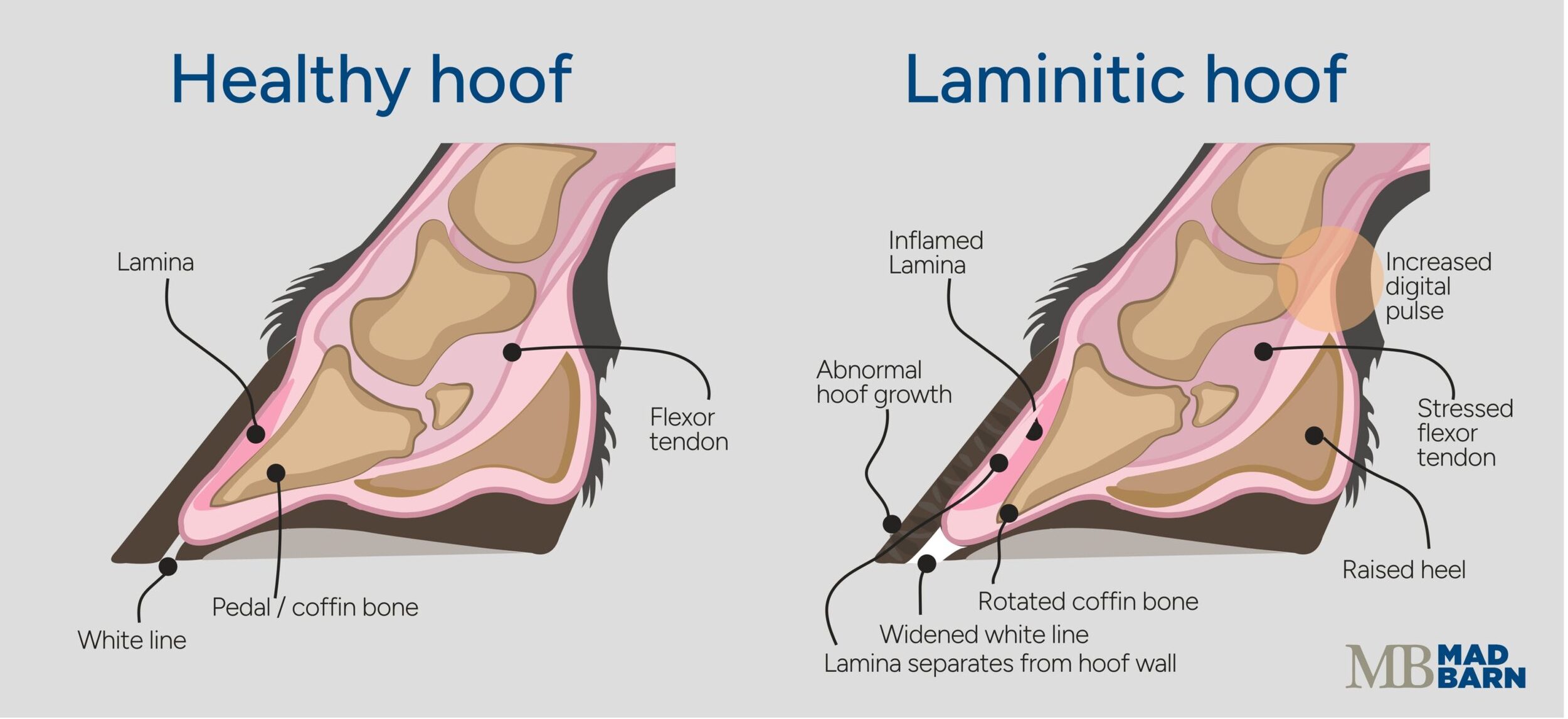 Illustration: Dr. Ana Mesa, PhD
The condition commonly affects more than one foot. Laminitis can also develop in a supporting limb when severe pain or injury in the opposite limb results in prolonged abnormal weight bearing. [14]
Affected horses may be reluctant to move and develop a short, careful gait. Other signs can include: [15]
- Repeated weight shifting
- An abnormal stance (“sawhorse” stance)
- Warm feet
- Strong digital pulses
Turning may be particularly uncomfortable because it increases uneven loading between the feet. [15]
 Sawhorse Stance Illustration: Dr. Ana Mesa, PhD
Laminitis is a medical emergency because damage within the hoof can progress and compromise the structural relationship between the hoof wall and coffin bone. Horses showing signs consistent with laminitis require prompt veterinary assessment rather than forced movement or a wait-and-see approach. [13][14][15]
Joint-Related Causes of Lameness in Horses
Joints allow movement while transferring substantial forces through the limbs. Pain can arise from several structures within or around a joint, including the cartilage, underlying bone, synovial membrane, joint capsule, and supporting ligaments. [16][17]
Joint pain may develop following acute injury or as part of a progressive condition. Depending on the structures involved, horses may show lameness, stiffness, reduced range of motion, joint swelling, or declining performance. [16][17][18]
Osteoarthritis
Osteoarthritis is a common cause of chronic or recurrent lameness in horses. It involves progressive changes within the joint, including deterioration of articular cartilage, changes to the underlying bone, and episodes of inflammation. [16][17]
Osteoarthritis can affect horses of any age and discipline, although previous injury, repetitive loading, conformation, and age may influence risk. [16][17]
Signs vary depending on the affected joint and stage of disease. Some horses are stiff after rest and improve as they warm up, while others become increasingly uncomfortable with exercise. [16][17]
Reduced range of motion, joint enlargement, or increased joint fluid may be present, but the absence of visible swelling does not rule out joint pain. [16][17]
Synovitis & Capsulitis
Synovitis is inflammation of the synovial membrane that lines a joint, while capsulitis refers to inflammation of the surrounding joint capsule. Factors contributing to inflammation in these tissues include: [16][17][19]
- Trauma
- Repetitive stress
- Instability
- Another joint disorder
Affected horses may show pain, swelling of the joint, stiffness, or reduced performance, and sometimes these signs occur before changes become visible on radiographs. [16][17][19]
Because synovitis and capsulitis can occur on their own or alongside other joint abnormalities, findings are best interpreted in the context of the entire joint and the horse’s workload. [16][17]
Osteochondritis Dissecans
Osteochondritis dissecans (OCD) is a developmental orthopedic disorder involving abnormal development of cartilage and the underlying bone. OCD is most commonly identified in young horses, although clinical signs may not become apparent until training begins and the affected joint is placed under greater load. [18]
Not every OCD lesion causes lameness. Some are incidental findings, while others contribute to joint swelling, stiffness, or pain. [18]
The clinical significance of an OCD lesion depends on factors including its location and size, the response of the affected joint, and the horse’s intended use. [18]
Septic Arthritis
Septic arthritis is an infection within a joint. Infection can develop following a penetrating wound, surgery, joint injection, or spread of bacteria through the bloodstream, particularly in foals. [20]
Although the presentation can vary, affected horses may develop: [20]
- Severe pain
- Heat
- Joint swelling
- Fever
Joint infection can rapidly damage articular cartilage and requires urgent veterinary treatment. Even a small wound near a joint warrants careful evaluation, as the joint capsule is still at risk of penetration despite minimal damage at the skin surface. [12][20]
Bone-Related Causes of Lameness in Horses
Bone injuries range from microscopic stress-related damage and bone bruising to complete fractures. The resulting lameness can vary considerably depending on the location, extent, and stability of the injury. [21]
Neither the amount of swelling nor the degree of lameness reliably indicates the severity of bone damage. Some significant injuries initially produce relatively subtle signs, making further investigation important when bone pain is suspected. [21][22]
Fractures
Complete or displaced fractures often cause sudden, severe lameness, but not every fracture is immediately obvious. [21][22]
Stable or incomplete fractures may allow a horse to continue bearing some weight on the affected limb. In contrast, abnormal limb angulation, instability, or sudden non-weight-bearing lameness raises concern for a more severe fracture. [21]
When a fracture is possible or suspected, owners should not exercise the horse to determine whether the lameness improves or becomes easier to identify. Additional movement can destabilize some injuries. [21][22]
Veterinary assessment before movement or transport is important because appropriate stabilization and handling depend on the location and type of fracture. [21][22]
Stress & Incomplete Fractures
Stress-related bone injuries develop when repeated loading exceeds the bone’s ability to adapt and repair. These injuries can occur during training as bone responds to repeated cycles of exercise. [22]
Early signs may be mild, intermittent, or only apparent during higher-intensity work. If loading continues, a small area of bone damage may progress to a more serious fracture. [22]
Early stress injuries are not always visible on radiographs. When clinical findings continue to suggest bone pain, horses may require repeat radiographs or advanced imaging such as nuclear scintigraphy, CT, or MRI. [22]
Bone Bruising
Bone bruising refers to microscopic injury and fluid changes within bone without a conventional fracture line. These changes may develop following direct impact or repetitive loading. [23]
Affected horses can develop persistent lameness even when radiographs appear normal. MRI can identify some forms of bone injury that are not visible with conventional radiography. [23][24]
Imaging findings still require interpretation alongside the clinical examination. Abnormalities identified on a scan do not always represent the primary source of the horse’s current lameness. [8][25]
Tendon & Ligament-Related Causes of Lameness in Horses
Tendons connect muscles to bones and transmit the forces generated by muscle contraction, while ligaments help stabilize joints and support the limb during movement. [26][27]
Injuries can occur suddenly following excessive loading or develop gradually through repeated strain. Acute injuries to superficial structures often cause heat, swelling, pain, and lameness, but deeper tendon and ligament injuries may produce few visible changes. [26][27][28]
Flexor Tendon Injuries
The superficial digital flexor tendon is commonly injured in athletic horses. Acute injury may cause warm, painful swelling along the back of the cannon region, with the degree of lameness varying according to the extent and location of the lesion. [26]
Ultrasound is commonly used to assess accessible tendon injuries, helping determine their location and extent. Repeat ultrasound examinations can also monitor healing and guide progression through rehabilitation. [29][30]
The deep digital flexor tendon extends down the back of the limb and into the hoof. Injuries within the hoof capsule can cause significant foot pain without visible swelling higher in the limb. [8]
Because veterinarians cannot fully assess these structures from the outside, diagnostic nerve blocks and advanced imaging such as MRI may be required to identify the source of pain. [8][10]
Suspensory Ligament Injuries
The suspensory apparatus supports the fetlock during weight bearing and helps control excessive extension of the joint. Injury can affect the upper portion of the suspensory ligament, its body, or either of its branches. [27]
Clinical signs vary with the location and severity of injury. Acute cases may produce localized pain and swelling, while chronic injuries can present primarily as reduced performance or persistent lameness with little obvious external change. [27]
Proximal suspensory pain in a hind limb can be particularly difficult to recognize because the resulting gait changes may resemble other hindlimb or back problems. [27]
Veterinarians typically combine physical examination, diagnostic nerve blocks, and imaging to localize the painful area and evaluate the affected structures. [8][27]
Collateral Ligament Injuries
Collateral ligaments help stabilize joints against excessive side-to-side movement. These ligaments are present at several locations throughout the limb, including within the hoof. [28]
Injury may follow a slip, sharp turn, or other episode of abnormal loading, although the resulting signs are not specific to collateral ligament damage. [28]
Ultrasound can evaluate accessible collateral ligaments. When the affected structure is located within the hoof capsule, MRI or other advanced imaging may help to identify and characterize the injury. [28]
Muscle, Back & Pelvis-Related Causes of Lameness in Horses
Musculoskeletal pain does not always originate in the lower limbs. Problems involving the muscles, back, sacroiliac region, or pelvis can also alter a horse’s gait and athletic performance. [3][8]
Direct trauma, slipping, overstretching, fatigue, or strenuous exercise can all injure muscles. A localized muscle injury can cause pain, swelling, weakness, or altered movement, while generalized muscle soreness may make a horse appear stiff or reluctant to move without producing a consistent limp. [31]
Exertional rhabdomyolysis is an important consideration when abnormal movement develops during or after exercise. This condition involves exercise-associated muscle damage and may cause painful or firm muscles, sweating, anxiety, and reluctance to move. [31]
Because exertional rhabdomyolysis can require specific treatment and management, affected horses should receive veterinary assessment rather than treatment for routine post-exercise stiffness. [31]
Pain involving the back, sacroiliac region, or pelvis can reduce impulsion, affect canter quality, cause resistance under saddle, or contribute to an uneven gait. Pelvic fractures and some other significant injuries may also be difficult to identify from external examination alone. [3][32]
Limb pain can contribute to secondary back soreness as the horse changes its movement to protect the affected area. Evaluating the horse as a whole helps distinguish primary pain from compensatory changes. [1][8][32]
Neurological & Mechanical Causes of Lameness in Horses
Not every abnormal gait is primarily due to musculoskeletal pain. Neurological disorders can affect the horse’s strength, coordination, and awareness of limb position, resulting in stumbling, toe dragging, abnormal foot placement, or other changes in movement. [1][25][33]
These abnormalities can resemble orthopedic lameness or occur at the same time as a painful musculoskeletal problem, which can make the underlying cause more difficult to determine. [25]
When neurological dysfunction is suspected, the veterinarian performs a neurologic examination to evaluate strength, coordination, awareness of limb position, and the horse’s response to controlled movements such as backing, turning, and navigating slopes. [25]
Mechanical lameness occurs when a physical restriction or abnormal movement alters gait without pain being the primary cause. Examples include some forms of restrictive scar tissue or abnormal movement of the patella. [34]
Although certain neurological or mechanical abnormalities can produce characteristic movement patterns, gait appearance is not always sufficient to determine the underlying cause. [33]
Trauma & Wound-Related Causes of Lameness
Trauma from kicks, falls, collisions, entanglement, or lacerations can damage structures ranging from the skin and muscles to bones, joints, tendons, and tendon sheaths. [6][20]
The appearance of a wound does not necessarily reflect the extent of the underlying injury. A relatively small puncture can penetrate a joint or tendon sheath and be more serious than a much larger superficial wound. [12][20]
A range of factors influence the significance of a wound, such as: [6][12][20]
- The location and depth of the injury
- Degree of contamination
- Bleeding
- Swelling
- Structures beneath the wound
What Causes Sudden Lameness in Horses?
Horses can become markedly lame within hours. Common causes of sudden lameness include: [1][8][35]
- A hoof abscess
- Sole bruising
- Lost or shifted shoes
- Penetrating injuries
- Acute tendon or ligament injuries
- Fractures
- Joint inflammation
- Laminitis
- Infection within a joint or tendon sheath
Sudden onset does not necessarily mean the injury occurred when the lameness was first noticed. [1]
A developing hoof abscess may become painful as pressure builds, while a stress injury may become apparent only after repeated loading. Chronic joint or soft-tissue problems can also flare suddenly. [7][36]
Serious injuries do not always cause swelling. Hoof pain, incomplete fractures, and injuries within the hoof capsule can cause significant lameness without obvious external signs. [21][22]
Owners should treat sudden non-weight-bearing lameness as potentially serious until a veterinarian determines the cause. [8][22]
Acute vs. Chronic Lameness
Lameness may be described as acute or chronic based on when it appeared and how it changes over time. These terms describe the pattern of lameness, not the exact cause, severity, or prognosis. [1][8]
Acute lameness develops suddenly. It may follow an obvious event, such as a kick, fall, slip, penetrating wound, or strenuous exercise, but it can also appear without a witnessed injury. [1][6][8]
Chronic lameness persists, recurs, or develops gradually over time. Because signs can develop slowly, owners may first notice reduced performance, stiffness, reluctance to work, or uneven movement rather than an obvious limp. [5][10][11][16]
Table 2. Acute and chronic lameness in horses
| Pattern | How It Appears | Common Causes | Important Consideration |
|---|---|---|---|
| Acute lameness |
|
|
|
| Chronic lameness |
|
|
|
The distinction between acute and chronic lameness provides useful context, but it does not replace diagnosis. Some serious injuries begin subtly, and some chronic problems worsen suddenly. A new or significant change in gait should not automatically be attributed to an existing diagnosis without reassessment. [1][16]
Determining where the problem originates is an important part of evaluating a lame horse. A veterinary lameness examination uses the horse’s history, physical examination, and gait assessment, with diagnostic blocks or imaging when needed, to localize the affected area and identify the underlying cause. [1][25]
Sudden, severe, or non-weight-bearing lameness requires prompt veterinary attention. When a serious injury such as a fracture is possible, unnecessary movement can worsen tissue damage and should be avoided until the horse is properly assessed. [1][21]
Signs of Lameness in Horses
Signs of lameness vary depending on the affected limb, the source of discomfort, and the severity of the underlying problem. Some horses develop an obvious limp, while others show subtle changes in stride, posture, willingness to move, or athletic performance. [1][2]
Common signs of lameness in horses include: [1][2][4][13]
- Uneven or shortened stride
- Head nodding or uneven movement of the pelvis
- Reluctance to bear weight on one limb
- Stiffness or difficulty turning
- Toe dragging or abnormal foot placement
- Reduced impulsion or willingness to move forward
- Difficulty with transitions or working on one rein
- Repeated refusal or reluctance at fences
- Heat, swelling, or pain in a limb or hoof
- Unexplained decline in performance
Subtle cases may show only one or two of these changes, making familiarity with the horse’s normal movement especially useful. [2][4]
Recognizing Subtle Lameness
Some cases of lameness only lead to subtle changes in movement. Subtle signs may first appear as changes in stride, stiffness, reduced impulsion, reluctance to move forward, difficulty on one rein, or an unexplained decline in performance. [1]
Lameness can be difficult to recognize when more than one limb is affected. If both front limbs or both hind limbs are painful, the horse may not consistently favor one leg. Instead, movement may appear as shortened strides, seem stiff, or restrict range of motion. [1][2]
These signs are not specific to lameness and can also occur with muscle weakness, neurological disease, or other problems affecting movement. A veterinary examination can help determine whether pain is involved, identify affected limbs or regions, and guide further investigation. [1][31]
When Lameness Is an Emergency
Some causes of lameness require urgent veterinary care because continued movement can worsen the injury or cause additional damage. Contact a veterinarian promptly if a horse has: [1][6][13]
- Sudden, severe, or non-weight-bearing lameness
- A limb that appears unstable, abnormally angled, or potentially fractured
- A penetrating hoof injury or embedded foreign object
- A wound over or near a joint, tendon sheath, tendon, or bone
- Marked or rapidly increasing swelling
- An abnormal stance, strong digital pulses, or other signs consistent with laminitis
- Fever, severe pain, distress, or rapidly worsening signs
Stop exercise and keep the horse as calm as possible without compromising safety. Contact your veterinarian before asking the horse to walk, attempting transport, or applying a splint. If a fracture or other unstable injury is present, unnecessary movement can cause further damage. [8][21][22]
If a nail or other object is embedded in the hoof, leave it in place until a veterinarian provides instructions. The location, depth, and angle of penetration may help determine which structures are affected. If the object has already come out, save it and photograph or mark the apparent entry site without probing or manipulating the foot. [6][12]
Not all cases of lameness in horses are emergencies. However, persistent, recurrent, or unexplained lameness still requires veterinary evaluation.
Early assessment can help identify the affected structure, limit further stress on injured tissues, and provide a clearer basis for treatment and rehabilitation. [1][3]
The Lameness Scale & Injury Severity
The severity of a horse’s lameness describes how apparent the gait abnormality is, but it does not necessarily indicate the severity of the underlying injury. [1][2]
Veterinarians commonly use the American Association of Equine Practitioners (AAEP) lameness grading scale to describe lameness in a consistent, standardized way. The scale ranges from Grade 0, where no lameness is detectable, to Grade 5, where the horse bears minimal weight on the affected limb or is unable to move.
Table 3. The AAEP lameness scale [8]
| Grade | Description |
|---|---|
| Grade 0 | Lameness is not perceptible under any circumstances. |
| Grade 1 | Lameness is difficult to observe and is not consistently apparent, regardless of circumstances. |
| Grade 2 | Lameness is difficult to observe at a walk or while trotting in a straight line but becomes consistently apparent under certain circumstances, such as circling, carrying weight, traveling on an incline, or moving over a hard surface. |
| Grade 3 | Lameness is consistently observable at the trot under all circumstances. |
| Grade 4 | Lameness is obvious at the walk. |
| Grade 5 | The horse bears minimal weight on the affected limb at rest or in motion, or is unable to move. |
The AAEP lameness grade describes how easily lameness is observed, not the amount of tissue damage or the horse’s prognosis.
A hoof abscess, for example, can cause marked lameness but improve relatively quickly with appropriate treatment. In contrast, some bone or soft-tissue injuries may initially cause only subtle lameness despite requiring a longer period of rehabilitation. [2]
For this reason, the lameness grade is only one part of the assessment. Identifying the affected structure and underlying cause is necessary to determine the significance of the problem and guide treatment. [1][2]
Factors That Can Increase Lameness Risk
Lameness can occur in any horse, including those with appropriate hoof care, conditioning, and management. However, certain factors can influence how forces are distributed through the limbs or affect how well bones, joints, tendons, ligaments, and muscles adapt to exercise. [26][27][31]
Factors that may increase the risk of lameness include: [1][3][5][36][27][31]
- Conformation: Limb alignment and hoof shape affect how weight and forces are distributed during movement. Certain conformational traits can place greater stress on specific structures, although conformation alone does not determine whether a horse will become lame.
- Hoof care: Overgrown feet, hoof imbalance, inadequate sole protection, and poorly fitted shoes can alter loading through the hoof and limb. Regular trimming or shoeing helps maintain appropriate hoof balance for the individual horse.
- Workload: Repetitive exercise or rapid increases in training intensity can place more stress on bone and soft tissues than they have adapted to tolerate. Gradual changes in workload allow the musculoskeletal system time to strengthen in response to exercise.
- Fitness: Fatigue can affect coordination and the horse’s ability to stabilize the limbs during movement. Conditioning should progress gradually and reflect the demands of the horse’s discipline and level of work.
- Footing: Hard, deep, uneven, rocky, or slippery surfaces can alter loading, increase concussion or fatigue, and affect traction. The effects of footing also depend on factors such as speed, workload, and the individual horse.
- Age: The types of musculoskeletal problems horses experience can change with age. Young horses are more commonly affected by developmental orthopedic conditions, while older horses are more likely to accumulate changes in joints and soft tissues.
- Previous injury: Previously injured tissues may respond differently to exercise, particularly if strength or function has not fully recovered. Horses may also change how they move in response to pain, increasing stress on other structures.
- Body condition: Excess body weight increases the mechanical load carried by the limbs. Inadequate energy, protein, or other nutrients can also affect muscle development, tissue repair, and the horse’s ability to meet the demands of exercise.
These factors often interact rather than acting independently. For example, a horse’s conformation may influence how it responds to a particular type of footing or workload, while fitness affects how well the musculoskeletal system tolerates those demands. [8][26][27]
Owners should tailor management to the individual horse. Appropriate conditioning, regular hoof care, suitable footing, adequate recovery, and balanced nutrition can help support musculoskeletal health across different ages and disciplines. [26][27][29]
How Lameness Is Diagnosed
A lameness examination aims to determine why a horse is moving abnormally and identify the structures involved. Because similar gait abnormalities can result from different conditions, accurately identifying the source of lameness can be difficult without a systematic veterinary examination. [8][35]
Involving a veterinarian is important when lameness is persistent, recurrent, unexplained, or severe. An accurate diagnosis helps guide treatment and rehabilitation while reducing the risk of continuing to exercise or manage the horse for the wrong underlying problem. [1][8][29]
The veterinarian first determines whether the abnormal movement is most consistent with pain, restricted movement, a neurological problem, or a combination of factors. When pain is suspected, the examination focuses on identifying the affected limb or region and localizing the source as precisely as possible. [1][25][33]
The diagnostic process typically combines the horse’s medical history, physical examination, and gait evaluation. Hoof testers, flexion tests, and diagnostic nerve or joint blocks may then help further localize pain. Once an area of concern is identified, diagnostic imaging is used to evaluate the structures within that region. [8]
Cases may not be diagnosed during a single examination. Subtle lameness, pain involving multiple areas, and some bone or soft-tissue injuries may require additional testing, advanced imaging, or reassessment over time. [8][19]
Medical History
A detailed history provides context for the examination and can help identify patterns or events associated with the onset of lameness.
The veterinarian typically asks when the change was first noticed, whether it developed suddenly or gradually, and whether the lameness is consistent or intermittent. [1][8]
Useful information can include: [1][3][8]
- Recent exercise, competition, turnout, travel, falls, or other trauma
- The gait, direction, surface, or movement that makes the lameness more apparent
- Changes in trimming, shoeing, tack, training, or workload
- Previous injuries, diagnostic findings, and treatments
- Rest, medications, or exercise performed since the signs began
- Fever, wounds, swelling, appetite changes, or other health concerns
Owners can also provide videos of the horse recorded when the problem is most apparent. This is particularly helpful when lameness is intermittent. However, owners should not recreate strenuous or unsafe conditions simply to obtain footage. [2][4][8]
Physical Examination
The physical examination helps identify visible or palpable abnormalities potentially associated with the horse’s gait. The veterinarian typically begins by observing the horse at rest, assessing posture, weight distribution, conformation, swelling, muscle loss, and signs of injury. [1][8]
The limbs may be palpated in both weight-bearing and non-weight-bearing positions to assess temperature, pain, joint filling, tissue contour, and range of motion. The veterinarian may also examine the feet, digital pulses, hoof walls, soles, shoes, neck, back, pelvis, and muscles. [1][5][8]
Comparing corresponding structures on opposite sides of the horse can help identify differences. However, natural asymmetry and pain affecting more than one limb can complicate interpretation.
Similarly, sensitivity during palpation does not necessarily mean that area is the primary source of lameness. [4][8][32][33]
Gait Evaluation
When it is safe to do so, the veterinarian observes the horse in motion. This can help identify the affected limb or region and recognize when the abnormality is most apparent. [35]
The horse is typically evaluated walking and trotting in a straight line on a firm, level, non-slip surface. The veterinarian assesses: [8]
- Rhythm
- Stride length
- Foot flight and placement
- Head movement
- Pelvic movement
- Weight bearing
Circles and different surfaces may make some abnormalities more apparent. Depending on the case, the veterinarian may also evaluate the horse while backing, turning, traveling on an incline, or moving under saddle. [8]
Owners should not trot or lunge a severely lame horse if they suspect a fracture or other unstable injury. In these cases, minimizing movement and evaluating the horse at rest take priority before further diagnostic procedures are considered. [8][22]
Veterinarians may also use sensor- or camera-based gait analysis to measure subtle movement asymmetries that are difficult to detect visually. These systems provide objective measurements but do not diagnose the cause of lameness. Results require interpretation alongside the rest of the clinical examination. [4]
Hoof Testers & Flexion Tests
Hoof testers and flexion tests can provide additional information about where pain may originate, but neither test identifies a specific injury on its own. [1][8]
Hoof testers apply controlled pressure to different areas of the hoof. A pain response can help the veterinarian identify discomfort within the foot, although hoof thickness, recent trimming, technique, and the horse’s individual sensitivity can affect the findings. [1][8]
During a flexion test, part of the limb is held in a flexed position for a set period before the horse trots away. The veterinarian then evaluates whether lameness temporarily becomes more apparent. [8]
Because flexion places stress on multiple structures at the same time, an increased response does not identify one specific structure or diagnosis. Instead, veterinarians interpret the result alongside the horse’s history, physical examination, and other findings. [1][8]
Nerve & Joint Blocks
Diagnostic analgesia, commonly called blocking, uses local anesthetic to temporarily reduce sensation in a selected area. This allows the veterinarian to determine whether pain from that region is contributing to the horse’s lameness. [8][19]
A nerve block desensitizes the region supplied by a particular nerve. A joint block places local anesthetic directly into a joint. [19]
If lameness improves after an area is desensitized, the result helps narrow down the source of pain. Blocks are often performed in a planned sequence, with the horse’s gait reassessed after each block. [8][19]
Results require careful interpretation because local anesthetic can spread to nearby tissues and some horses have more than one source of pain. Diagnostic blocks are also not appropriate for every case and are generally avoided when an unstable fracture is suspected. [8][19]
Diagnostic Imaging
Diagnostic imaging is generally most informative after the clinical examination has localized the area of concern. Different imaging methods provide information about different tissues, so the veterinarian selects the technique based on the structures and conditions being investigated. [8][12]
Some of these methods include: [8][12][22][26][27][28][30]
- Radiographs (X-rays): Provide images of bone, joint margins, alignment, and some structures within the hoof. Radiographs are commonly used to investigate fractures, arthritis, and changes associated with laminitis. However, early stress injuries and many soft-tissue injuries may not be visible.
- Ultrasound: Uses sound waves to evaluate accessible tendons, ligaments, muscles, joint margins, and fluid-filled structures such as joints and tendon sheaths. Ultrasound is also used to monitor tissue healing during rehabilitation.
- MRI: Magnetic resonance imaging provides detailed images of both bone and soft tissues. It is particularly useful for investigating structures within the hoof that are difficult to evaluate with other imaging methods.
- CT: Computed tomography produces detailed cross-sectional images and is particularly useful for evaluating complex bony anatomy. Equipment and the ability to image standing horses vary between facilities.
- Nuclear scintigraphy (“bone scan”): This technique identifies areas of altered bone activity and may help locate regions of concern when pain is difficult to localize or more than one site is suspected.
Veterinarians may recommend additional testing when the examination suggests a problem that musculoskeletal imaging alone cannot fully evaluate. Bloodwork may help investigate systemic illness or muscle disease, and veterinarians can analyze fluid from a joint or tendon sheath when they suspect infection.
A neurological examination may be appropriate when weakness, poor coordination, or abnormal limb placement suggests nervous system involvement. [25][31][33]
Reaching a diagnosis may require more than one examination or type of diagnostic test. This is particularly true when lameness is subtle, more than one area is painful, or an injury is difficult to identify with initial imaging. [8][19]
Treatment of Lameness in Horses
Treatment for lameness depends on the underlying cause. Because lameness is a clinical sign rather than a specific disease, identifying the affected structure and type of injury is important before developing a treatment plan. [1][8]
Management may vary based on the severity and duration of the injury, the tissues involved, the horse’s intended use, and the risk of further damage. Depending on the diagnosis, treatment may include: [1]
- Rest or controlled exercise
- Pain management
- Therapeutic farriery
- Veterinary procedures
- Surgery
- Physical rehabilitation
Rest is not sufficient to address every cause of lameness, and complete inactivity is not appropriate for every stage of healing. Bone, tendon, ligament, cartilage, and muscle respond differently to both loading and rest.
Rehabilitation therefore aims to protect injured tissues while gradually restoring movement and function as healing progresses. [1][29]
Rest & Controlled Exercise
The appropriate amount of movement during recovery depends on the injury and stage of healing. Some acute injuries initially require stall rest or confinement to limit loading, while other conditions may allow limited turnout or controlled hand walking. [29][30]
As healing progresses, owners can gradually introduce controlled exercise. An equine rehabilitation program may progress from walking to trotting, strengthening exercises, and eventually a return to the horse’s normal workload. [29]
Progress is based on the horse’s comfort, veterinary reassessment, and evidence of tissue healing rather than a fixed timeline alone. A horse may appear more comfortable before the injured structure has regained sufficient strength to tolerate normal work. [29][30]
Increasing exercise too quickly can overload healing tissues and increase the risk of reinjury. However, prolonged inactivity can also contribute to loss of fitness and muscle strength. The goal is to provide an appropriate level of loading for the tissue and stage of recovery. [30]
Pain & Inflammation Management
Managing pain and inflammation can improve comfort during the early stages of some injuries and support an appropriate rehabilitation plan. [1]
Veterinarians may recommend cold therapy for certain acute injuries to help manage heat, swelling, and pain. The method, frequency, and duration depend on the affected tissue and diagnosis. [37]
Horses may also require non-steroidal anti-inflammatory drugs (NSAIDs) and other medications to manage pain and inflammation. Because these medications can cause adverse effects and may mask pain without resolving the underlying injury, owners should use them only under veterinary guidance. [15][16]
Improved comfort after medication does not necessarily mean an injured structure has healed. A horse should not return to work solely because medication masks lameness. [15][16]
Farriery & Hoof Support
Trimming and shoeing can influence the distribution of force through the hoof and lower limb. Changes in hoof balance, breakover, weight distribution, or sole protection may therefore form part of the treatment plan for some causes of lameness. [5][11]
The best approach depends on the underlying condition and whether the hoof is involved. Horses with sole bruising, laminitis, tendon injuries, joint disorders, or hoof imbalances require different types of hoof support. [10][15]
Radiographs can show the position and alignment of the coffin bone within the hoof capsule, as well as changes in sole depth, hoof wall thickness, and bone structure. These findings can help guide decisions about trimming, shoe placement, and other forms of hoof support. [8][10][11][15]
Hoof care may also need to be adjusted during rehabilitation as the foot grows and the horse’s movement and workload change. Communication between the veterinarian and farrier can help ensure the approach remains appropriate throughout recovery. [10][11][29]
Joint-Directed & Other Veterinary Treatments
Depending on the diagnosis, veterinarians may recommend additional treatments to manage pain, inflammation, or tissue injury. These can include: [16][29][30]
- Medications administered into or around a joint
- Biological therapies
- Shockwave therapy
- Therapeutic exercise
- Other rehabilitation techniques
The evidence supporting individual therapies varies by treatment and condition. A therapy that is appropriate for one type of injury may provide little benefit for another. [16][20][30]
For this reason, these treatments are generally most useful when selected for a specific diagnosis and incorporated into a broader management and rehabilitation plan. They do not replace appropriate exercise management, rehabilitation, or hoof care. [16][20][30]
Surgery
Veterinarians may recommend surgery when an injury cannot be adequately managed with conservative treatment or when surgical intervention offers the best opportunity to restore function or control disease. [20][22][30]
Conditions that may require surgery include: [18][22]
- Certain fractures
- Infected joints or tendon sheaths
- Penetrating injuries
- Osteochondritis dissecans (OCD) fragments
- Selected tendon or ligament injuries
Depending on the condition, surgery may stabilize a fracture, remove damaged or abnormal tissue, treat infection, or restore function to an affected structure. [22][30]
Whether surgery is appropriate depends on the location and severity of the lesion, timing of treatment, available facilities, the horse’s intended use, and expected quality of life. Postoperative care and rehabilitation also play an important role in recovery. [29][30]
Return to Work
Returning a horse to work is a gradual process because improvement in lameness does not necessarily mean the affected tissue has fully healed. Different tissues regain strength at different rates, and their ability to tolerate exercise may continue to improve after the horse appears comfortable. [29]
Workload is typically increased in stages as healing progresses. A horse may be comfortable walking before the affected structure is ready for trotting, circles, jumping, speed work, or abrupt changes in direction. [29][30]
Veterinary reassessment can help determine when exercise can safely progress. Depending on the injury, this may include evaluating gait, pain, swelling, or changes on diagnostic imaging. [29][30]
The goal of rehabilitation depends on the individual horse and diagnosis. Some horses return to their previous level of performance, while others may require a less demanding workload or ongoing management to maintain long-term comfort and function. [29][30]

Prognosis for a Lame Horse
The prognosis for a lame horse depends primarily on the underlying diagnosis rather than how obvious the lameness appears. [1][2][8]
Important factors include the structure involved, the type and extent of tissue damage, how long the condition has been present, whether infection or instability is involved, and how the horse responds to treatment and rehabilitation. Age, intended use, and the demands of the horse’s discipline can also influence the expected outcome. [8][27][30]
Recovery times vary considerably between causes of lameness. A straightforward hoof abscess or minor soft-tissue injury may resolve relatively quickly with appropriate treatment. Significant tendon, ligament, bone, or joint injuries can require several months or longer for tissues to heal and regain sufficient strength for exercise. [27][29][30]
It is also important to distinguish between different measures of recovery. Comfort at rest, pasture soundness, return to light exercise, and return to previous athletic performance are separate outcomes. A horse may be comfortable and have a good quality of life without returning to its former level of work, while another may return to athletic activity but require ongoing management. [22][27][29][30]
Prognosis may become clearer as the horse progresses through treatment and rehabilitation. Follow-up examinations and, when appropriate, repeat diagnostic imaging can help the veterinarian assess healing and determine whether the horse is ready for increasing levels of exercise. [8][27][29][30]
Supporting Musculoskeletal Health
Lameness episodes in horses are not always preventable. However, consistent management can help reduce avoidable stress on the musculoskeletal system, support tissue adaptation to exercise, and make changes in movement easier to recognize. [27][29]
Helpful management steps to reduce the risk of lameness include: [8][26][38][27][29]
- Maintain regular hoof care: Schedule trimming or shoeing according to the horse’s hoof growth, conformation, workload, and individual needs. Regularly check shoe fit, hoof balance, cracks, thrush, and sole condition between appointments.
- Build fitness gradually: Increase exercise duration, intensity, speed, and technical difficulty in stages. Progressive conditioning gives bone, muscle, tendons, and ligaments time to adapt to increasing workloads. Owners should also incorporate appropriate warm-up, cool-down, and recovery periods into training.
- Manage footing: Check riding and turnout areas for holes, rocks, slippery surfaces, and inconsistent footing depth. Hard, deep, or unstable footing can alter loading and increase fatigue or loss of traction, so workload may need adjustment when conditions change.
- Maintain a healthy body condition: Excess body weight increases the mechanical load placed on the limbs. At the same time, inadequate energy or protein intake can limit muscle development, tissue repair, and recovery from exercise.
- Feed a balanced diet: Provide adequate forage through a forage-first feeding program that meets the horse’s requirements for energy, protein, vitamins, and minerals. These nutrients support muscle function, bone health, and tissue repair. Supplements should complement a balanced diet rather than compensate for deficiencies in the foundation feeding program.
- Monitor normal movement: Become familiar with the horse’s usual gait, posture, feet, and limb contours. Changes in stride, willingness to move, or performance may be early signs of discomfort and require investigation if they persist or recur.
Management should be individualized according to the horse’s: [27][29][30]
- Age
- Health
- Discipline
- Conformation
- Workload
- Previous injuries
Horses returning from injury may require additional adjustments to exercise, footing, hoof care, and recovery as their tissues gradually regain the capacity to tolerate normal loading. [27][29][30]
Veterinarians, farriers, qualified equine nutritionists, and rehabilitation professionals can work together to address different factors that influence musculoskeletal health, particularly in horses with complex or recurrent lameness.
Frequently Asked Questions
Here are some frequently asked questions about lameness in horses:
The most common causes of lameness in horses include hoof problems, joint disorders, bone injuries, tendon or ligament damage, muscle pain, and trauma. Hoof abscesses, sole bruising, laminitis, osteoarthritis, fractures, and soft-tissue injuries are frequently encountered. Some abnormal gaits can also result from neurological disease, mechanical restrictions, or muscular disorders rather than musculoskeletal pain. The likely cause depends on the horse's age, workload, history, and clinical signs.
Sudden lameness in horses can be caused by a hoof abscess, sole bruise, lost or shifted shoe, penetrating hoof injury, acute tendon or ligament injury, fracture, joint inflammation, laminitis, or infection within a joint or tendon sheath. The underlying problem may have developed before the lameness became noticeable, as can happen when pressure builds inside a hoof abscess or repeated loading worsens a stress injury. Sudden, severe, or non-weight-bearing lameness requires prompt veterinary attention.
The first signs of lameness in a horse may include an uneven or shortened stride, stiffness, head nodding, uneven pelvic movement, toe dragging, or difficulty turning. Reduced impulsion, reluctance to move forward, difficulty with transitions, and an unexplained decline in performance can also indicate discomfort. Early or intermittent lameness may be subtle and may not produce an obvious limp.
A horse may be suddenly lame without swelling when the pain originates inside the hoof or another deep structure. Hoof abscesses, sole bruises, incomplete fractures, and injuries to structures enclosed within the hoof capsule can cause significant pain with few visible external changes. A lack of swelling does not rule out a serious injury, so sudden or severe lameness should be assessed promptly by a veterinarian.
You can help determine which leg a horse is lame on by observing stride length, weight bearing, head movement, and pelvic movement. With forelimb lameness, the horse commonly raises its head when the painful limb bears weight. Hindlimb lameness may produce uneven movement of the pelvis or a reduced push from the affected leg. Subtle lameness, compensatory movement, or pain affecting multiple limbs can make the source difficult to identify without a veterinary examination.
A horse can be lame without showing an obvious limp, particularly when the pain is mild, intermittent, or present in more than one limb. Subtle signs can include stiffness, a shortened stride, reduced impulsion, resistance under saddle, difficulty on one rein, reluctance to work, or declining performance. Similar changes can also occur with weakness, neurological disease, or other movement disorders, so persistent or recurrent signs warrant veterinary evaluation.
Lameness in a horse is an emergency when it is sudden, severe, or non-weight-bearing, or when a fracture, laminitis, penetrating injury, or infection is possible. Other warning signs include limb instability, abnormal angulation, fever, severe pain, rapidly increasing swelling, strong digital pulses, or a wound near a joint or tendon sheath. Stop exercise, keep the horse as quiet as safely possible, and contact a veterinarian before walking, transporting, or attempting to splint the horse.
Whether you should walk a lame horse depends on the cause and severity of the lameness. A horse with severe or unexplained acute lameness should not be exercised or walked to see whether the problem improves. If a fracture or unstable injury is possible, unnecessary movement can cause further damage. Controlled walking may be introduced during rehabilitation, but only according to veterinary guidance and the stage of tissue healing.
Veterinarians diagnose lameness in horses by combining the horse's history with a physical examination and gait evaluation. Hoof testers, flexion tests, and diagnostic nerve or joint blocks may help localize the painful area. Radiographs, ultrasound, magnetic resonance imaging, computed tomography, or nuclear scintigraphy can then be selected according to the structures being investigated. Subtle or complex cases may require more than one examination or type of diagnostic test.
Lameness in horses is treated according to the underlying cause, the affected tissue, and the risk of further injury. Treatment may include rest, controlled exercise, pain management, therapeutic hoof care, veterinary procedures, surgery, or a structured rehabilitation program. Rest alone is not appropriate for every condition or every stage of healing. The treatment plan should protect damaged tissues while gradually restoring movement, strength, and function.
How long lameness takes to heal depends on the diagnosis, the tissues involved, and the extent of the damage. A straightforward hoof abscess or minor soft-tissue injury may improve relatively quickly with appropriate treatment, while significant tendon, ligament, bone, or joint injuries can require several months or longer. Recovery should be guided by comfort, veterinary reassessment, and evidence of tissue healing rather than a fixed timeline. A horse may appear sound before the injured structure is ready for normal work.
A horse can recover completely from some causes of lameness, but the expected outcome depends on the diagnosis, severity of tissue damage, treatment, rehabilitation, and intended use. Some horses return to their previous level of athletic performance, while others remain comfortable with a reduced workload or ongoing management. Comfort at rest, pasture soundness, return to light exercise, and return to full performance are different measures of recovery. Follow-up examinations can help determine when the horse is ready for increased activity.
Owners can reduce the risk of lameness in horses through regular hoof care, gradual conditioning, suitable footing, balanced nutrition, and appropriate recovery between workouts. Maintaining a healthy body condition and adjusting exercise to the horse's fitness, conformation, age, and previous injuries can also help limit excessive strain. Owners should become familiar with the horse's normal gait, posture, feet, and limb contours so subtle changes can be recognized early. Not every case is preventable, but persistent or recurrent changes in movement should be investigated.
Summary
The causes of lameness in horses range from common hoof problems to serious injuries and disorders affecting bones, joints, soft tissues, muscles, or the nervous system. Accurate diagnosis is essential because the appearance of lameness does not always reflect the severity of the underlying condition.
- Hoof abscesses, bruising, laminitis, shoeing problems, and punctures are common causes
- Joint disease, fractures, bone bruises, and soft-tissue injuries can cause mild or severe signs
- Back pain, muscle disorders, neurological issues, and mechanical restrictions can alter gait
- Non-weight-bearing lameness, fractures, puncture wounds, and laminitis signs need urgent care
- Veterinarians use history, exams, diagnostic blocks, and imaging to localize the source
- Treatment may include controlled exercise, pain management, farriery, rehabilitation, or surgery
References
- Adams. S. B. Overview of Lameness in Horses. Merck Veterinary Manual. 2025.
- Dyson. S. Can Lameness Be Graded Reliably?. Equine Veterinary Journal. 2011. View Summary .
- Boado. A. et al. Orthopaedic Injuries in 272 Dressage Horses: A Retrospective Study. Animals. 2025. View Summary .
- Hardeman. A. M. et al. Visual Lameness Assessment in Comparison to Quantitative Gait Analysis Data in Horses. Equine Veterinary Journal. 2022. View Summary .
- Mata. F. et al. Investigating Associations between Horse Hoof Conformation and Presence of Lameness. Animals. 2024. View Summary .
- Smith. M. R. W. Penetrating Injuries of the Foot. Equine Veterinary Education. 2013.
- Redding. W. R. and O'Grady. S. E. Septic Diseases Associated with the Hoof Complex: Abscesses, Punctures Wounds, and Infection of the Lateral Cartilage. Veterinary Clinics of North America: Equine Practice. 2012. View Summary .
- Baxter. G. M. et al. Adams and Stashak's Lameness in Horses. 7th ed. Wiley. 2020.
- Beasley. B. Bruising of the Hoof Capsule in Horses. Merck Veterinary Manual. 2024.
- Redding. W. R. and O'Grady. S. E. Nonseptic Diseases Associated with the Hoof Complex: Keratoma, White Line Disease, Canker, and Neoplasia. Veterinary Clinics of North America: Equine Practice. 2012. View Summary .
- Hunt. R. J. Farriery for the Hoof with Low or Underrun Heels. Veterinary Clinics of North America: Equine Practice. 2012. View Summary .
- Michotte. M. et al. Diagnostic Accuracy of Plain Radiography to Identify Synovial Penetration in Horses with Traumatic Limb Wounds. Equine Veterinary Journal. 2025. View Summary .
- Wylie. C. E. et al. Decision-Tree Analysis of Clinical Data to Aid Diagnostic Reasoning for Equine Laminitis: A Cross-Sectional Study. Veterinary Record. 2016. View Summary .
- van Eps. A. et al. Supporting Limb Laminitis. Veterinary Clinics of North America: Equine Practice. 2021.
- Mitchell. C. F. et al. The Management of Equine Acute Laminitis. Veterinary Medicine: Research and Reports. 2015. View Summary .
- Goodrich. L. R. and Nixon. A. J. Medical Treatment of Osteoarthritis in the Horse - A Review. The Veterinary Journal. 2006. View Summary .
- McIlwraith. C. W. et al. Biomarkers for Equine Joint Injury and Osteoarthritis. Journal of Orthopaedic Research. 2018. View Summary .
- Martinez-Saez. L. et al. Osteochondrosis in Horses: An Overview of Genetic and Other Factors. Equine Veterinary Journal. 2026. View Summary .
- Radtke. A. et al. Intra-Articular Anaesthesia of the Equine Stifle Improves Foot Lameness. Equine Veterinary Journal. 2020. View Summary .
- de Souza. T. C. et al. A Scoping Review of the Current Evidence on Treatment and Outcomes Following Synovial Sepsis. Equine Veterinary Journal. 2022. View Summary .
- Wright. I. M. Racecourse Fracture Management. Part 2: Techniques for Temporary Immobilisation and Transport. Equine Veterinary Education. 2017.
- Bowers. K. et al. A Review of Equine Tibial Fractures. Equine Veterinary Journal. 2023. View Summary .
- Olive. J. et al. Use of standing low-field magnetic resonance imaging to diagnose middle phalanx bone marrow lesions in horses. Equine Veterinary Education. 2009.
- Powell. S. E. et al. Standing magnetic resonance imaging detection of bone marrow oedema-type signal pattern associated with subcarpal pain in 8 racehorses: A prospective study. Equine Veterinary Journal. 2010.
- Blythe. L. L. and Engel. H. N. Neuroanatomy and Neurological Examination. Veterinary Clinics of North America: Equine Practice. 1999. View Summary .
- Thorpe. C. T. et al. A Review of Tendon Injury: Why Is the Equine Superficial Digital Flexor Tendon Most at Risk?. Equine Veterinary Journal. 2010. View Summary .
- Guest. D. J. et al. A Review of the Equine Suspensory Ligament: Injury Prone yet Understudied. Equine Veterinary Journal. 2025. View Summary .
- Lamb. L. et al. Clinical Outcome of Collateral Ligament Injuries of the Tarsus. The Canadian Veterinary Journal. 2012. View Summary .
- Davidson. E. J. Controlled Exercise in Equine Rehabilitation. Veterinary Clinics of North America: Equine Practice. 2016. View Summary .
- Smith. R. K. W. Treatment of Tendinopathies. Equine Veterinary Education. 2024.
- Valberg. S. J. Sporadic and Recurrent Exertional Rhabdomyolysis. Veterinary Clinics of North America: Equine Practice. 2025. View Summary .
- le Jeune. S. and Jones. J. Prospective Study on the Correlation of Positive Acupuncture Scans and Lameness in 102 Performance Horses. American Journal of Traditional Chinese Veterinary Medicine. 2014.
- Saville. W. J. A. et al. Interobserver Variation in the Diagnosis of Neurologic Abnormalities in the Horse. Journal of Veterinary Internal Medicine. 2017. View Summary .
- Dumoulin. M. et al. Upward Fixation of the Patella in the Horse: A Retrospective Study. Veterinary and Comparative Orthopaedics and Traumatology. 2007. View Summary .
- Rodrigues. A. P. C. et al. Objective Lameness Assessment of 235 Horses Undergoing Lameness Examination in Brazil: A Retrospective Study. Brazilian Journal of Veterinary Medicine. 2025. View Summary .
- Birch. H. L. et al. Macroscopic 'Degeneration' of Equine Superficial Digital Flexor Tendon Is Accompanied by a Change in Extracellular Matrix Composition. Equine Veterinary Journal. 1998. View Summary .
- 10.1016/j.jevs.2009.10.019.
- O'Brien. C. et al. Microdamage in the Equine Superficial Digital Flexor Tendon. Equine Veterinary Journal. 2021. View Summary .