
 English
Hock fusion occurs when the bones in one of the horse’s lower hock joints gradually grow together, becoming a solid structure without distinct edges. It most commonly develops in horses with osteoarthritis of the lower hock, also known as bone spavin.
Horses with lower hock arthritis may develop stiffness, hind limb lameness or changes in performance. As the condition progresses, the affected joint can fuse completely. Once this happens, pain associated with arthritis often improves because the affected joint can no longer move.
Although a fused joint sounds concerning, the lower hock joints normally have very little movement. Many horses remain comfortable and participate in riding and competition once hock fusion is complete.
Treatment focuses on managing pain and keeping the horse comfortable. In selected cases, veterinarians may also consider procedures to encourage fusion when lower hock pain persists despite other treatments.
What is Hock Fusion in Horses?
Bone fusion is the gradual joining of the bones of a joint until movement between them is reduced or eliminated. [1][2] In horses, hock fusion usually occurs in lower hock structures as osteoarthritis progresses.
The ends of bones are normally covered by smooth articular cartilage and separated by a small joint space containing synovial fluid. Together, these structures allow bones to move smoothly against one another, giving range of motion to the limb. [2]
When osteoarthritis develops, inflammation in the joint leads to degenerative cartilage damage and changes on the bone surfaces. Over time, the joint space may narrow and new bone can form around and across the affected joint, merging separate structures into a single mass of bone. [2]
At first, only part of the joint may be bridged by new bone. This is known as partial fusion, and some joint space and movement may remain. [2]
With complete fusion, the joint space is lost and the bones become permanently joined. In horses with painful distal tarsal osteoarthritis, complete fusion is associated with improved lameness and comfort. [2][3]
Hocks affected by arthritis do not always progress to complete fusion. Some joints remain partially fused for long periods or never fuse completely.
Anatomy of the Hock
The horse’s hock, or tarsus, contains several bones and joints that support the hind limb and allow movement.
Most of the visible bending and straightening of the hock occurs in the upper tarsocrural joint. The joints below it have much less movement. The two lower joints most commonly affected by osteoarthritis and fusion are the distal intertarsal joint and the tarsometatarsal joint. [4]
Because these joints normally move very little, complete fusion can eliminate a source of pain without greatly reducing the range of motion in the affected hock.
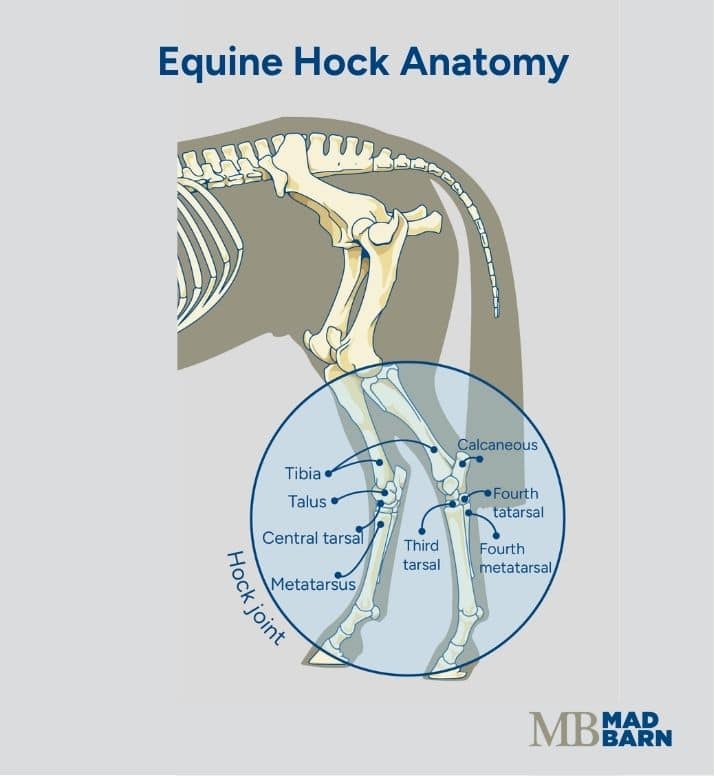 Illustration: Dr. Ana Mesa, PhD
What's your top priority with your horse's health?

What's your top priority with your horse's health?
Signs of Hock Fusion in Horses
Horses with fusing hocks may show stiffness, hind limb lameness or changes in performance. The signs are subtle at first and become more noticeable as osteoarthritis progresses. [1]
Some horses develop obvious lameness, while others merely seem less comfortable using their hindquarters. Owners may first notice the horse takes longer to loosen up, has less impulsion or does not perform certain movements as easily as before.
Common signs of hock fusion in horses include:
- Hind limb stiffness
- Subtle or intermittent lameness
- Shortened hind limb stride
- Reduced impulsion
- Difficulty engaging the hindquarters
- Stiffness when beginning exercise
- Reduced willingness to move forward
Some horses appear stiff when they first begin moving and get more comfortable as they warm up. This pattern is not specific to hock arthritis, and similar signs can occur with other causes of hind limb pain.
The severity of discomfort can change over time. A horse may have periods of increased stiffness or lameness followed by periods when they appear more comfortable. These changes do not indicate whether the hock has fused or how far the process has progressed.
Changes Under Saddle
Owners often notice changes under saddle due to hock arthritis before obvious lameness develops. Riders may feel the horse is less willing to push from behind or has more difficulty with movements that require greater use of the hindquarters. [1]
Changes under saddle include:
- Difficulty with transitions
- Reduced ability or willingness to collect
- Difficulty picking up or maintaining a canter lead
- Resistance to lateral work
- Difficulty backing
- Reduced jumping performance
- Reluctance to engage the hindquarters
These signs are not specific to hock fusion and can occur with many musculoskeletal conditions. Other signs that warrant veterinary assessment include:
- Persistent stiffness
- Lameness
- Changes in performance
When to Call Your Veterinarian
Contact your veterinarian if your horse develops new or persistent hind limb lameness, increasing stiffness or an unexplained decline in performance.
Veterinary assessment is especially important if you notice:
- Sudden or significant lameness
- Increasing difficulty moving or exercising
- Heat or swelling around the hock
- Increasing reluctance to engage the hindquarters
- Pain that is no longer controlled with the current treatment plan
- Recurring lameness after a period of improvement
There are many possible causes of hind limb lameness, and signs that appear to come from the hock may originate elsewhere. An accurate diagnosis directs treatment at the source of pain.
Causes of Hock Fusion in Horses
Hock fusion is most commonly associated with bone spavin, or osteoarthritis of the lower hock joints. A combination of joint loading, conformation, workload and individual susceptibility can contribute to wear and inflammation over time. [5][6]
Factors that contribute to lower hock osteoarthritis include:
- Repetitive loading of the lower hock joints
- Hind limb conformation
- Type and intensity of athletic work
- Previous injury or trauma
- Age and accumulated joint stress
- Individual risk factors for osteoarthritis
- Obesity
In particular, repetitive movements that increase concussion, rotational forces, or weight-bearing stress place greater demands on the lower hock joints and increase the risk of bone spavin.
Examples include collected movements in dressage, takeoffs and landings in show jumping, sliding stops and spins in reining, tight turns in barrel racing, and repeated high-speed impacts in racing. [3]
Lower hock osteoarthritis is more common in older horses due to cumulative wear and tear on the joints. However, this condition can affect younger horses, particularly when conformation, development, previous injury or workload increases stress on these joints.
Diagnosis of Hock Fusion in Horses
Diagnosing hock fusion involves determining whether the lower hock joints are a source of pain and assessing the changes occurring within them.
Veterinarians typically combine the horse’s history, physical examination, lameness evaluation, diagnostic analgesia and imaging findings to reach a diagnosis.
Lameness Examination
A lameness exam begins with observing the horse standing and moving to look for asymmetry, stiffness or other abnormalities.
Horses with lower hock osteoarthritis may show reduced hock movement, shortened forward movement of the hindlimb, or dragging of the hind toe.
Depending on the horse and severity of lameness, the examination can include movement in a straight line and on a circle, sometimes over different surfaces.
A veterinarian may also palpate the hocks and surrounding structures for swelling, heat, pain or bony enlargement.
Hock Flexion Tests
The examination may include a flexion test, in which the veterinarian holds the limb flexed briefly before assessing the horse at a trot. [7]
An increase in lameness after hock flexion supports suspicion of lower hock pain, but flexion test results must be interpreted by a veterinarian alongside a lameness examination. [7]
Diagnostic Analgesia (Nerve & Joint Blocks)
Diagnostic analgesia, sometimes referred to as a nerve block, involves temporarily numbing a specific region or joint to localize the origin of pain. [5]
If lameness improves significantly after the veterinarian numbs the lower hock, this region is likely involved in the initiation of pain. [5]
Radiography (X-Rays)
Radiographs, more commonly known as X-rays, are typically used to evaluate horses with suspected lower hock osteoarthritis and to assess the progression of fusion. [2] Abnormalities visible on X-ray do not always correspond directly with the location or severity of a horse’s lameness. [5]
An X-ray can identify changes in your horse’s joints including:
- Narrowing or loss of the joint space
- Changes in the bone beneath the cartilage
- New bone formation around the joint
- Irregular joint margins
- Bony bridging across the joint
As fusion advances, the normal space between adjacent bones becomes increasingly difficult to distinguish. Complete bony bridging indicates the joint has fused. [2]
Radiographic findings are interpreted together with the horse’s clinical signs because the severity of visible changes does not always match the degree of pain. [5] Once hock fusion is detected, the treating veterinarian may recommend regular follow-up imaging to monitor the extent of fusion and the presence of other bony changes.
Advanced Imaging
The veterinarian may recommend more detailed imaging when the source of lameness remains unclear or standard radiographs do not provide enough information.
Computed tomography (CT) or nuclear scintigraphy (“bone scan”) may be useful in selected cases. The most appropriate test depends on the individual horse and the suspected source of pain. [2]
Treatment During Hock Fusion
Treatment of hock function focuses on controlling pain and inflammation while keeping the horse as comfortable and active as their condition allows.
The best treatment plan depends on which joints are affected, the severity of lameness, the extent of joint changes, how the horse responds to intervention and the horse’s intended use.
Joint Injections
Veterinarians may recommend intra-articular medication for horses with painful lower hock osteoarthritis. [8]
Corticosteroids help reduce inflammation within affected joints, and the veterinarian may select additional medications based on the horse’s needs and examination findings. [8]
Joint injections are intended to manage inflammation and improve comfort. They do not stop or accelerate the fusion process. [8]
Anti-Inflammatory Medication
Non-steroidal anti-inflammatory drugs (NSAIDs) reduce the pain and inflammation associated with hock arthritis in horses. [9]
The choice of medication, dose and duration depend on the individual horse. Discuss long-term or repeated use with your veterinarian because NSAIDs are associated with adverse side effects in horses, particularly when used incorrectly. [9]
Exercise, Rehabilitation & Supportive Care
Appropriate movement and conditioning are important parts of managing osteoarthritis, but exercise recommendations should match the horse’s comfort and stage of disease. [10]
Some horses remain in regular work with appropriate treatment. Others need reduced or modified exercise during painful periods. A significantly lame horse requires evaluation before owners attempt to increase exercise in an effort to encourage fusion.
Owners should provide their horse with regular hoof care to help maintain hoof balance and reduce strain on the lower hock joints. Your veterinarian and farrier may recommend changes to trimming or shoeing based on the horse’s conformation, movement and individual needs.
Natural vs. Induced Hock Fusion
Lower hock joints can gradually develop bony bridging as osteoarthritis progresses. In some horses, this may eventually result in complete fusion, or ankylosis, of the affected joint. [11]
When lower hock pain persists despite appropriate treatment, veterinarians may consider procedures intended to encourage complete fusion. This is known as induced arthrodesis. [12]
The goal of induced arthrodesis is to eliminate movement within the painful low-motion joint. Once the bones have successfully fused, movement through that joint stops. Discomfort associated with the damaged joint surfaces may improve.
Induced fusion is not appropriate for every horse with bone spavin. Before considering the procedure, the veterinarian must determine which joint is causing the horse’s pain and whether it is an appropriate candidate for arthrodesis. [12]
Surgical Arthrodesis
Surgical arthrodesis uses techniques that disrupt or remove the remaining articular cartilage and encourage bone formation across the joint. [13][14]
Veterinarians use several surgical approaches to address the lower hock joints. The technique chosen depends on the affected joint, severity of disease, and the veterinary surgeon’s recommendation. [13][15]
Fusion does not occur immediately after surgery. Bone must gradually bridge the joint during recovery. Horses require an appropriate rehabilitation and pain-management program while healing takes place. [13][14]
Complications are possible and should be discussed with the veterinary surgeon before treatment.
Chemical Arthrodesis
Chemical arthrodesis involves introducing a substance into an affected joint to damage the remaining articular cartilage and encourage fusion. [16]
Alcohol has been used to induce hock fusion in horses with lower hock osteoarthritis. Because the substance can damage healthy cartilage if it reaches an unintended joint, careful assessment of the affected joint and any communication with neighboring joints is essential before treatment. [16][12]
Recovery & Prognosis
The prognosis for hock fusion depends largely on which joint is affected, whether fusion remains partial or progresses to completion, and whether the horse has other sources of lameness.
Recovery time varies depending on whether fusion occurs naturally or is induced. Veterinary rechecks and follow-up radiographs can assess how fusion is progressing and help guide rehabilitation and return to work.
After Complete Hock Fusion
Many horses have reduced pain and improved lameness scores after complete fusion of a painful lower hock joint. [13]
Because these joints contribute relatively little to overall hock movement, some horses can return to their previous level of athletic activity after appropriate rehabilitation. Others may require a modified workload depending on the severity of their original joint disease and presence of other orthopedic problems. [13][3]
Riding Horses with Fused Hocks
Many horses can be ridden after successful fusion of the lower hock joints if they are comfortable and otherwise sound. [3][13]
A fused lower hock does not automatically mean a horse must retire from riding. Suitability for a particular discipline or workload depends on the individual horse’s soundness, overall musculoskeletal health, and veterinary assessment.
Fusion involving a high-motion joint has different implications and is not equivalent to fusion of the lower hock joints.
Persistent Lameness After Fusion
Complete fusion of an affected joint should eliminate movement within that joint, but it does not resolve every source of hind limb pain. The horse may have osteoarthritis in another hock joint or pain originating elsewhere in the limb or body. Long-standing lameness can also alter how a horse moves and loads other structures.
If a horse remains lame after documented hock fusion, further veterinary evaluation can help identify other possible sources of discomfort.

Managing a Horse With Hock Arthritis
Management of a horse affected by hock arthritis should not aim to accelerate joint fusion. Instead, it should focus on:
- Keeping the horse comfortable
- Maintaining appropriate activity
- Supporting long-term soundness
Regular veterinary reassessment can help determine whether the horse’s comfort or treatment needs change over time. Exercise should match the horse’s current level of soundness, and conditioning should progress gradually with modifications during more painful periods.
Consistent hoof care is important. Appropriate trimming or shoeing can help maintain hoof balance and reduce unnecessary mechanical stress on the hind limbs. Maintaining a healthy body condition can further reduce the load placed on arthritic joints.
Owners should monitor the horse’s usual movement, behavior and willingness to work. Early indications that a horse is less comfortable include:
- Stiffness
- Shortened hind limb stride
- Reduced impulsion
- Changes in performance
Nutrition & Body Condition
Horses affected by fused hocks or arthritis benefit from a carefully formulated diet to maintain appropriate body condition and supply essential nutrients involved in maintaining joint health.
Keeping your horse at a healthy body condition helps prevent excessive loading of the joints. Excess body weight increases mechanical stress on arthritic hocks. Regular conditioning and adequate muscle development support joint stability and normal movement.
Nutrition influences joint function and overall soundness by supplying the nutrients required to maintain articular cartilage, connective tissues, synovial fluid, bones, muscles and hooves.
A balanced diet should provide adequate energy, protein, essential fatty acids, vitamins and minerals to support musculoskeletal health. These nutrients are required for muscle maintenance, cartilage and connective tissue metabolism, bone mineralization and healthy hoof growth, all of which play a role in soundness. Adequate vitamin and mineral intake also supports normal cellular function, antioxidant defenses and tissue repair.
Horses who need additional joint support often benefit from supplementation with long-chain omega-3 fatty acids. Eicosapentaenoic acid (EPA) and docosahexaenoic acid (DHA) are omega-3s that are incorporated into cell membranes and used by the body to produce compounds that regulate inflammation and promote its resolution.
Mad Barn’s OmegaPrime+ provides 7,500 mg of combined DHA and EPA per 30 mL serving, along with 750 IU of vitamin E, an antioxidant that helps protect cells from oxidative damage.

Other joint supplements may provide additional support for joint health, but the effectiveness of individual ingredients varies. When selecting a supplement, look for ingredients supported by equine research and supplied at physiologically relevant doses.
Read our guide to the best joint supplements for horses for a research-based comparison of ingredients, their benefits and effective doses.
Frequently Asked Questions
Here are some frequently asked questions about hock fusion in horses:
When a horse's hocks fuse, new bone grows across an affected joint until the bones on either side become permanently joined. Fusion most commonly affects the lower hock joints in horses with osteoarthritis, also known as bone spavin. Once fusion is complete, movement within that joint is eliminated, which may relieve pain from the damaged joint surfaces.
Horses' hocks usually fuse as a consequence of chronic osteoarthritis in the lower hock joints. Damage to the cartilage causes changes in the underlying bone, narrowing of the joint space and new bone formation around the joint. Over time, this new bone may bridge the joint and permanently join the adjacent bones.
The hock joints most likely to fuse are the distal intertarsal and tarsometatarsal joints. These lower joints normally contribute very little to the bending and straightening of the hock. Fusion of a higher-motion joint is a different clinical problem and can have a much greater effect on movement and athletic function.
Hock fusion can be painful while osteoarthritis is active and the affected joint remains partially mobile. During partial fusion, movement can continue between damaged joint surfaces and cause ongoing discomfort. Once a low-motion joint has fused completely, pain arising from movement within that joint may improve substantially or resolve.
Signs associated with fusing hocks can include hindlimb stiffness, intermittent lameness, a shortened stride, reduced impulsion and difficulty engaging the hindquarters. Under saddle, a horse may struggle with transitions, collection, canter leads, lateral work, backing or jumping. These signs can also occur with other musculoskeletal conditions, so they cannot confirm that fusion is taking place.
Hock fusion is diagnosed using the horse's history, a physical examination, a lameness evaluation and imaging findings. Flexion tests and diagnostic analgesia may help determine whether the lower hock is an important source of pain. Radiographs can show narrowing of the joint space, new bone formation and bony bridging, but the findings must be interpreted alongside the horse's clinical signs.
The time required for a horse's hocks to fuse varies with the affected joint, the severity of osteoarthritis and whether fusion occurs naturally or is induced. Natural fusion is unpredictable, and some joints remain partially fused for long periods or never fuse completely. Induced fusion also requires time for new bone to bridge the joint, followed by appropriate rehabilitation and veterinary monitoring.
A horse's lower hock joints may fuse naturally as osteoarthritis progresses, a process known as ankylosis. However, not every arthritic joint will progress to complete fusion, and the timing cannot be reliably predicted. Management should focus on controlling pain, maintaining appropriate activity and supporting soundness rather than relying on eventual fusion as the only treatment strategy.
A veterinarian may encourage hock fusion through induced arthrodesis when pain from a lower hock joint persists despite appropriate treatment. Surgical procedures can disrupt or remove remaining cartilage, while chemical arthrodesis uses a substance to damage the cartilage and promote fusion. These procedures are only appropriate for selected horses after the painful joint and any communication with neighbouring joints have been carefully assessed.
Whether a horse can be ridden while its hocks are fusing depends on its comfort, degree of lameness, stage of disease and veterinary recommendations. Some horses can continue suitable exercise with treatment, while others need a reduced or modified workload during painful periods. A significantly lame horse should be evaluated before exercise is increased or changed.
A horse can often be ridden after successful fusion of the lower hock joints if it is comfortable and otherwise sound. Because these joints normally contribute relatively little to overall hock movement, complete fusion does not automatically require retirement from riding. The appropriate discipline and workload depend on the horse's rehabilitation, overall musculoskeletal health and veterinary assessment.
A completely fused joint generally does not require injections to control pain within that joint because it can no longer move. However, another hock joint may still be affected by osteoarthritis and require treatment. Persistent or recurring lameness should be evaluated rather than assumed to come from the fused joint.
A horse may remain lame after its hocks have fused if another joint or structure is still causing pain. Osteoarthritis may affect a different hock joint, or discomfort may originate elsewhere in the limb or body. Long-standing lameness can also change how the horse moves and loads other structures, so further veterinary evaluation may be needed.
A horse with fusing hocks should be managed with a plan focused on comfort, appropriate activity and long-term soundness. Treatment may include joint medication, non-steroidal anti-inflammatory drugs, modified exercise, rehabilitation and individualized hoof care under veterinary guidance. Maintaining a healthy body condition and monitoring changes in stiffness, stride, behaviour and willingness to work can also support ongoing management.
A veterinarian should examine a horse with new or persistent hindlimb lameness, increasing stiffness or an unexplained decline in performance. Prompt assessment is especially important when lameness is sudden or significant, the hock is hot or swollen, movement is becoming more difficult or the current treatment no longer controls the pain. An accurate diagnosis is essential because signs that appear to involve the hock may originate elsewhere.
Complete hock fusion cannot be reversed because bone has permanently bridged the joint. Joint supplements and other treatments cannot restore movement once the bones have fused. However, fusion of a lower hock joint does not necessarily limit a horse's usefulness, and many horses remain comfortable and active after successful fusion.
Summary
Hock fusion most commonly develops as osteoarthritis causes progressive bony bridging across one of the horse's low-motion lower hock joints. Complete fusion can relieve joint pain, although diagnosis, treatment and prognosis depend on the affected joint and the horse's overall soundness.
- Partial fusion may remain painful because movement continues within the portion of the joint that has not fused
- Signs can include hindlimb stiffness, intermittent lameness, reduced impulsion and difficulty engaging the hindquarters
- Diagnosis combines the horse's clinical history, physical and lameness examinations, diagnostic analgesia and imaging findings
- Treatment may include joint injections, anti-inflammatory medication, appropriate exercise, rehabilitation and supportive hoof care
- Selected horses with persistent lower hock pain may undergo surgical or chemical arthrodesis to encourage complete fusion
- Many horses can return to riding after complete lower hock fusion if they are comfortable, otherwise sound and appropriately rehabilitated
References
- Khumsap. S. et al. Effect of Induced Unilateral Synovitis of Distal Intertarsal and Tarsometatarsal Joints on Sagittal Plane Kinematics and Kinetics of Trotting Horses. American Journal of Veterinary Research. 2003.
- Steiger. J. I. et al. Diagnostic Performance of Radiography for the Evaluation of Osteoarthritis in the Equine Distal Tarsus: Comparison with Computed Tomography. Animals. 2025.
- Sanchez-Arjona. R. et al. Outcomes of Diode Laser-Facilitated Ankylosis of the Distal Tarsal Joints in Horses: A Retrospective Study of 64 Cases. Veterinary Surgery. 2026.
- Sprackman. L. et al. Relationship between the Shape of the Central and Third Tarsal Bones and the Presence of Tarsal Osteoarthritis. The Veterinary Journal. 2015.
- Byam-Cook. K. L. and Singer. E. R. Is There a Relationship between Clinical Presentation, Diagnostic and Radiographic Findings and Outcome in Horses with Osteoarthritis of the Small Tarsal Joints?. Equine Veterinary Journal. 2009.
- Eksell. P. et al. Prevalence and Risk Factors of Bone Spavin in Icelandic Horses in Sweden: A Radiographic Field Study. Acta Veterinaria Scandinavica. 1998.
- Björnsdóttir. S. et al. Radiographic and Clinical Survey of Degenerative Joint Disease in the Distal Tarsal Joints in Icelandic Horses. Equine Veterinary Journal. 2000.
- McIlwraith. C. W. The Use of Intra-Articular Corticosteroids in the Horse: What Is Known on a Scientific Basis?. Equine Veterinary Journal. 2010.
- Jacobs. C. C. et al. Non-Steroidal Anti-Inflammatory Drugs in Equine Orthopaedics. Equine Veterinary Journal. 2022.
- Gough. M. R. et al. Tiludronate Infusion in the Treatment of Bone Spavin: A Double Blind Placebo-Controlled Trial. Equine Veterinary Journal. 2010.
- Barneveld. A. Spavin in Horses. Tijdschrift voor Diergeneeskunde. 1990.
- Lamas. L. P. et al. Use of Ethanol in the Treatment of Distal Tarsal Joint Osteoarthritis: 24 Cases. Equine Veterinary Journal. 2012.
- Adkins. A. R. et al. Surgical Arthrodesis of Distal Tarsal Joints in 17 Horses Clinically Affected with Osteoarthritis. Australian Veterinary Journal. 2001.
- Dechant. J. E. et al. Use of a Three-Drill-Tract Technique for Arthrodesis of the Distal Tarsal Joints in Horses with Distal Tarsal Osteoarthritis: 54 Cases (1990-1999). Journal of the American Veterinary Medical Association. 2003.
- Zubrod. C. J. et al. Comparison of Three Methods for Arthrodesis of the Distal Intertarsal and Tarsometatarsal Joints in Horses. Veterinary Surgery. 2005.
- Carmalt. J. L. et al. Alcohol-Facilitated Ankylosis of the Distal Intertarsal and Tarsometatarsal Joints in Horses with Osteoarthritis. Journal of the American Veterinary Medical Association. 2012.